The Control of Porcine Circovirus Diseases (PCVDs)

Main objectives


These objectives are listed in the order of their importance to the project.

To apply the information generated to the elimination and/or control of PCVD.

To initiate and maintain a proactive information dissemination programme aimed at all relevant stakeholders, including consumers, producers and policy makers.

To identify the common co-factors/triggers in epizootic PCVD scenarios necessary for the full development of clinical disease and evaluate the importance of air-born spread of PCVD/PMWS.

To determine the role of nutrition in the susceptibility/resistance to PCVD.

To determine the sites of replication of PCV2 and early pathogenesis of PCVDs.

To elucidate the early interactions of PCV2 virus with the host immune system.

To elucidate the role of porcine genetics in susceptibility/resistance to PCVD.

To determine the molecular processes of PCV2 replication (18 months) and virulence.

To standardise and harmonise and distribute reagents, and SOPs for use within the consortium. 


Contact us


“The Control of Porcine Circovirus Diseases (PCVDs): Towards Improved Food Quality and Safety” research programme started on December 1, 2004 and will run for 51 months. It has been funded by the EU Sixth Framework Programme.

Porcine Circovirus

EM Porcine Circovirus 2 (QUB)The goal of this project is to better understand the role of Porcine Circovirus in diseases of pigs. It will generate information on control measures that will have a positive impact on the health and welfare of pigs. It will also help producers meet consumer concerns for quality and safety of pork products. The lead organisation is Queen's University Belfast and there are 16 partners from EU and North America. The consortium combines the existing strengths of the partners from two previous projects in EU Framework 5, with expertise in epizootiology, nutrition, porcine genetics, bacteriology and information dissemination.

Sixth Framework Logotype

Project No.: 513928

Sixth Framework Programme

Priority SSP/5.4.6

(Priority 5. Food Quality and Safety)

Copyright © 2005 - 2010 Meat and Livestock Commission, UK | HTML 4.01 | CSS


 Background of the proposal


The members of this consortium have had an active research programme on PCVDs for the last 7 years and their collective research findings and publications in peer reviewed journals (> 100) represent the major advances in PCVD research and the state of the art.


Porcine circovirus:


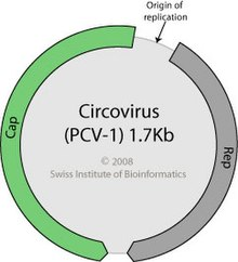
Porcine circovirus (PCV) was first identified in 1974 as a contaminant of the continuous pig kidney cell line PK/15 [63]. This virus was later shown to contain a single-stranded, circular DNA genome. A "novel" PCV-like virus was first isolated from pigs with a wasting disease in western Canada in 1998 [17]. Shortly thereafter, similar viruses were isolated from diseased pigs in N America and Europe [1, 2, 57]. These isolates were shown to be antigenically and genomically distinct from PCV isolates and were designated PCV2 viruses, to discern from the previous virus, which was named PCV1 [8]. PCV1 and PCV2 are small (17nm) icosahedral, non-enveloped viruses containing a single-stranded, circular DNA genome and are now classified in the circovirus genus of the family Circoviridae [24, 25, 60, 61, 63, 64]. They show an ambisense genome organisation with 2 major open reading frames for replication and packaging of viral DNA [34, 35, 36, 38, 39, 40, 41, 42, 62]. The exact mechanisms of viral replication and interaction with host factors are still not known. Mechanisms of PCV2 replication and the molecular basis of pathogenesis of the virus will be addressed in Work Package 4 of this project.


Field disease:


A wasting syndrome in Canadian and French pigs was first reported in 1996 and named postweaning multisystemic wasting syndrome (PMWS) [13, 33]. PCV nucleic acid and antigen were demonstrated in abundance in the lesions of affected pigs and subsequent isolation and characterisation of a PCV2 virus from diseased pigs was reported [1, 2, 17, 53]. Since these initial reports of PCV2-associated wasting disease in piglets in Canada and France the disease has been reported in almost all pig producing countries around the world [37, 49, 57]. Gross lesions of PCVD/PMWS include generalised lymphadenopathy, hepatitis, nephritis and pneumonia and typical histological lesions include lymphocytic depletion together with histiocytic and multinucleated giant cell infiltration in lymph nodes, degeneration and necrosis of hepatocytes, and multifocal lymphohistiocytic interstitial pneumonia [1, 26, 53]. The criteria used for the diagnosis of PCVD/PMWS include the existence of compatible clinical signs, presence of characteristic microscopic lesions in lymphoid tissues and detection of PCV2 within these lesions [59]. Reagents and SOPs for diagnosis and detection of PCVD/PMWS will be optimised and harmonised in Work Package 1 of this project.


PCV2 is widespread in pigs throughout the world and retrospective analyses of sera from 1969 onwards have shown the presence of antibody to a PCV2 virus in a high percentage of the sera tested [14, 43, 44, 51, 52, 65]. Retrospective analyses of tissue sections from diseased pigs has shown that sporadic cases of classical PCVD/PMWS have occurred as far back as the early 1986 [51, 55]. Evidence is emerging that PCV2 may play a major role in other porcine disease syndromes, including proliferating and necrotising pneumonia [1,48], reproductive disorders in pigs [31, 66] and porcine dermatitis and nephropathy syndrome (PDNS) [1, 20, 59]. PDNS was first described in S America in 1976, but until recently occurred only sporadically in EU member states. However, outbreaks of PDNS over the past 5 years have spiralled to epidemic proportions in EU member states and elsewhere. A recent survey in UK identified 251 cases (9.6 % of larger pig holdings). In many of these incidents, PDNS progressed from a sporadic to an epizootic form, with a case mortality of 25 to 30%. Mortalities can reach 100%, as has been reported for PDNS outbreaks in Spain. The microscopic lesions of PDNS are indicative of an immune complex-mediated disease, typically those of a type III hypersensitivity reaction. Nevertheless, epidemiological evidence suggests that PDNS is an infectious disease. The emergence of epidemic PDNS in recent years parallels the appearance of PCVD/PMWS leading to speculation that this syndrome is also PCV2-related [45]. However, to date, no consistent model of experimental production of PDNS has been developed. This will be addressed in Work Package 5 of this project. PCV2 is not a new virus and PCVD/PMWS is not a new disease. It is not known why sporadic PCVD/PMWS has emerged during the last decade as a global epizootic. Information generated in Work Package 2 of this project will be important in answering this, and other questions related to PCVD/PMWS epizootiology.


Experimental infections:


Clinical disease, and gross and histological lesions consistent with PCVD/PMWS have been reproduced following experimental infection of gnotobiotic, colostrum-deprived (CD) and colostrum-fed (CF) piglets with PCV2 [3, 4, 5, 6, 7, 9, 10, 11, 12, 18, 21, 22, 26, 28, 29, 30, 50, 54]. Consistent reproduction of clinical disease in an experimental model seems to require PCV2 infection plus modulation of the immune system by either co-infection with other viruses (porcine parvovirus (PPV)/porcine reproductive and respiratory disease virus (PRRSV) or the use of non-infectious immune modulators [3, 4, 5, 22, 26, 28, 29, 30, 50]. Recent studies using a gnotobiotic model have shown that inoculation of pigs with PCV2 alone plus a non-specific stimulation of immune system results in clinical PCVD/PMWS in 100% of the inoculates [28]. To date, this remains the only 100% disease model, which uses PCV2 as the only infectious agent. However, reproducible clinical disease can be achieved by the co-inoculation of CD pigs with PCV2 and PPV [3, 7], or cloned PCV2 DNA with PPV [unpublished]. Also experimental in-utero infection of porcine foetuses has resulted in gross and histological lesions in the inoculated foetuses, that vary in severity, dependent on the state of gestation of the foetus when inoculated [56]. Stimulation of the immune system of young pigs in current husbandry practises can be multifactorial and PCVD/PMWS is now considered as a multifactorial syndrome where PCV2 is essentially. Mechanisms of PCV2 pathogenesis will be elucidated in Work Packages 5 and 6 of this project.


Immunology and Immunopathogenesis:


Infected pigs seroconvert to PCV2, however the specific roles of the different immune compartments in protection against disease is unknown [27]. Studies have shown that PCV2 accumulates in macrophages and dendritic cells. This is likely to be a pivotal event in the pathogenesis of the disease. In-vivo studies on tissues from PMWS-affected pigs have shown that macrophages and/or dendritic cells contain large amounts of PCV2 antigen [18, 28, 29, 53]. Recent in-vitro studies have confirmed that PCV2 does not replicate in these cell types [19]. However, importantly, PCV2 infectivity was not reduced following infection of macrophages and in-vitro culture for up to 8 days. In vitro studies have revealed potentially immunoregulatory sequences in the genome of PCV2 that inhibits induction of IFN-alpha production [23].


Field and experimental studies have shown significant changes in the subpopulations of blood peripheral mononuclear cells of diseased pigs. These changes were characterised by lymphopenia, increase of circulating monocytes, reduction of T cells (mainly CD4+ and/or CD8+, as well as double positive cells) and B lymphocytes when compared with clinically healthy, non-PCV2 infected pigs [16, 47, 58]. In addition, more recent work have shown cytokine mRNA alterations in PMWS affected pigs, which were characterized by an over expression of IL-10 mRNA in thymus and IFN-gamma mRNA in tonsils, and by decreases of several cytokines in other lymphoid tissues [15].


All together, these immunopathological findings in PMWS affected pigs suggest an inability to mount an effective immune response. However, it still remains unclear how this virus acts upon the immune system of infected pigs, especially during the early phases of infection. This will be addressed in Work Packages 5 and 6 of this project.


Control of PCVDs:


Currently very little is known about control of PCVDs. Recent field studies have indicated that a "20-point plan" of improved husbandry measures may sometimes, but not always, limit the disease impact in affected herds [32]. Importantly, field observations by veterinarians and producers suggest that susceptibility/resistance to PCVD/PMWS may be influenced by the genetics or breed of the host, specifically in regard to the boar lines used. None of these “observations” have been scientifically evaluated or assessed. This will be addressed in Work Package 3 of this project. In addition, on some farms the use of feed additives and alterations in feeding regimes have had a beneficial effect on PCVD/PMWS. However, on other farms this has had no effect. This will be addressed in Work Package 7 of this project. To date, no commercial vaccines are available for PCVD. Strategies for the control of PCVDs will be developed through the collaborative efforts of all partners and particularly through data obtained from Work Packages 2, 3, 5, 6, 7 and 8. Information generated within all of the above Work Packages will be disseminated through Work Package 9 of the project.


Summary:


Since the initiation of the two research projects on PCVD under Framework 5, a great deal of basic information on the pathogenesis, epidemiology and replication of PCV2 has been generated and a new generation of biological and procedures for the study of this disease have been prepared. This information and these biological and procedures will serve as a sound platform for further research in this proposed project by an enhanced new multidisciplinary consortium. The new consortium combines the existing strengths of the partners from the two previous projects with expertise in epizootiology, nutrition, porcine genetics, bacteriology and information dissemination.


References:


Albina E, Truong C, Hutet E, Blanchard P, Cariolet R, L'Hospitalier R, Mahe D, Allee C, Morvan H, Amenna N, Le Dimna M, Madec F and Jestin A. An experimental model for post-weaning multisystemic wasting syndrome (PMWS) in growing piglets. Journal of Comparative Pathology 2001; 125: 292-303.

Allan GM, Phenix K, Todd D and McNulty M. Some biological and physico-chemical properties or porcine circovirus. Journal of Veterinary Medicine B 1994; 41: 17-26.

Allan GM, McNeilly F, Foster JC, Adair BM. Infection of leucocyte cell cultures derived from different species with pig circovirus. Veterinary Microbiology 1994; 41(3):267-279.

Allan GM, Mackie DP, McNair J, Adair BM, McNulty MS. Production, preliminary characterisation and applications of monoclonal antibodies to porcine circovirus. Veterinary Immunology and Immunopathology 1994 Nov; 43(4):357-371.

Allan GM, McNeilly F, Cassidy JP, Reilly GA, Adair B, Ellis WA, McNulty MS. Pathogenesis of porcine circovirus; experimental infections of colostrum deprived piglets and examination of pig foetal material. Veterinary Microbiology 1995; 44(1):49-64.

Allan GM, McNeilly F, Kennedy S, Daft B, Clarke EG, Ellis JA, Haines DM, Meehan BM, Adair BM. Isolation of porcine circovirus-like viruses from pigs with a wasting disease in the USA and Europe. Journal of Veterinary Diagnostic Investigation 1998; 10(1):3-10.

Allan G, Meehan B, Todd D, Kennedy S, McNeilly F, Ellis J, Clark EG, Harding J, Espuna E, Botner A, Charreyre C. Novel porcine circoviruses from pigs with wasting disease syndromes. Veterinary Record 1998; 142(17):467-468.

Allan GM, McNeilly F, Meehan BM, Kennedy S, Mackie DP, Ellis JA, Clark EG, Espuña E, Saubi N, Riera P, Botner A and Charreyre CE. Isolation and characterisation of circoviruses from pigs with wasting syndromes in Spain, Denmark and Northern Ireland. Veterinary Microbiology 1999; 66: 115-123.

Allan GM, Kennedy S, McNeilly F, Foster JC, Ellis JA, Krakowka SJ, Meehan BM and Adair BM. Experimental reproduction of severe wasting disease by co-infection of pigs with porcine circovirus and porcine parvovirus. Journal of Comparative Pathology 1999; 121: 1-11.

Allan GM, McNeilly F, Meehan BM, Ellis JA, Connor TJ, McNair I, Krakowka S, Kennedy S. A sequential study of experimental infection of pigs with porcine circovirus and porcine parvovirus: immunostaining of cryostat sections and virus isolation. Journal of Veterinary Medicine B Infect Dis Vet Public Health 2000; 47(2):81-94.

Allan GM, McNeilly F, Kennedy S, Meehan B, Ellis J, Krakowka S. Immunostimulation, PCV-2 and PMWS. Veterinary Record 2000; 147(6):170-171.

Allan GM, McNeilly E, Kennedy S, Meehan B, Moffett D, Malone F, Ellis J and Krakowka S. PCV-2-associated PDNS in Northern Ireland in 1990. Porcine dermatitis and nephropathy syndrome. Veterinary Record 2000; 146:711-712.

Allan GM, McNeilly F, Ellis J, Krakowka S, Meehan B, McNair I, Walker I, and Kennedy S. Experimental infection of colostrum deprived piglets with porcine circovirus 2 (PCV2) and porcine reproductive and respiratory syndrome virus (PRRSV) potentiates PCV2 replication. Archives of Virology 2000; 145: 2421-2429.

Allan GM and Ellis JA. 2000. Porcine circoviruses: a review. Journal of Veterinary Diagnostic Investigation; 12: 3-14.

Allan GM, McNeilly F, McNair I, Curran MD, Walker I, Ellis J, Konoby C, Kennedy S, Meehan B. Absence of evidence for porcine circovirus type 2 in cattle and humans, and lack of seroconversion or lesions in experimentally infected sheep. Archives of Virology 2000; 145(4):853-857.

Allan GM, McNeilly F, McNair I, O’Connor M, Meehan B, Gilpin D, Ellis J, Townsend H, Lasagna C, Boriosi G and Krakowka S. Neonatal vaccination for Mycoplasma hyopneumoniae and post-weaning multisystemic wasting syndrome: a field trial. Pig Journal 2001; 48: 34-41.

Allan GM, McNeilly F, Meehan B, Kennedy S, Johnston D, Ellis J, Krakowka S, Fossum C, Wattrang E and Wallgren P. Reproduction of PMWS with a 1993 Swedish isolate of PCV-2. Veterinary Record 2002; 150: 255-256.

Allan GM, McNeilly F, McNair I, Meehan B, Marshall B, Ellis J, Lasagna C, Boriosi G, Krakowka S, Reynaud G, Boeuf-Tedeschi L, Bublot N and Charreyre C. Passive transfer of maternal antibodies to PCV2 protects against development of post-weaning multisystemic wasting syndrome (PMWS) experimental infections and a field study. Pig Journal 50:59-67.

Allan G, McNeilly F, Meehan B, McNair I, Ellis J, Krakowka S, Fossum C, Wattrang E, Wallgren P, Adair B. Reproduction of postweaning multisystemic wasting syndrome in pigs experimentally inoculated with a Swedish porcine circovirus 2 isolate. Journal of Veterinary Diagnostic Investigation 2003; 15(6):553-560.

Baekbo P, Hassing AG, Olsen P, Lorenzen B, Lauridsen C. Vitamin E status of the weaned pig as a riskfactor for dying of PMWS. Proceedings of the 4th International Symposium on Emerging and Re-emerging Pig Diseases. PRRS, PMWS, Swine Influenza, Rome 2003: 220-221.

Balasch M, Segalés J, Rosell C, Domingo M, Mankertz A, Urniza A and Plana-Durán J. Experimental inoculation of conventional pigs with tissue homogenates from pigs with post-weaning multisystemic wasting syndrome. Journal of Comparative Pathology 1999; 121:139-148.

Beattie, V.E., Burrows, M.S., Moss, B.W., Weatherup, R.N. The effect of food deprivation prior to slaughter on performance, behaviour and meat quality. Meat Science 2002; 62: 412-418.

Beattie, V.E., Weatherup, R.N., Kilpatrick, D.J. The effect of providing additional feed in a highly accessible trough on feeding behaviour and growth performance of weaned pigs. Irish Journal of Agricultural and Food Research 1999; 38: 209-216.

Beattie, V.E., Weatherup, R.N., Moss, B.W., Walker, N. The effect of increasing carcass weight of finishing boars and gilts on joint composition and meat quality. Meat Science 1999; 52:205-212.

Blanchard P, Mahe D, Cariolet R, Truong C, Le Dimna M, Arnauld C, Rose N, Eveno E, Albina E, Madec F, Jestin A. An ORF2 protein-based ELISA for porcine circovirus type 2 antibodies in post-weaning multisystemic wasting syndrome. Veterinary Microbiology 2003; 94(3):183-194.

Blanchard P, Mahe D, Cariolet R, Keranflec'h A, Baudouard MA, Cordioli P, Albina E, Jestin A. Protection of swine against post-weaning multisystemic wasting syndrome (PMWS) by porcine circovirus type 2 (PCV2) proteins. Vaccine 2003; 21(31):4565-4575.

Brunet S., and Charreyre C. Comparison of sequences of PCV2 strains, Proceedings of the 16th IPVS – Melbourne Australia 2000; 629

Bogdan J, West K, Clark E, Konoby C, Haines D, Allan G, McNeilly F, Meehan B, Krakowka S, Ellis JA. Association of porcine circovirus 2 with reproductive failure in pigs: a retrospective study, 1995-1998. Canadian Veterinary Journal 2001; 42(7):548-550.

Calsamiglia M, Segales J, Quintana J, Rosell C and Domingo M. Detection of Porcine Circovirus Types 1 and 2 in Serum and Tissue Samples of Pigs with and without Postweaning Multisystemic Wasting Syndrome. Journal of Clinical Microbiology 2002; 40:1848-1850.

Carrasco L, Segalés J, Bautista MJ, Gómez-Villamandos JC, Rosell C, Ruiz-Villamor E and Sierra MA. Intestinal chlamydial infection concurrent with postweaning multisystemic wasting syndrome in pigs. Veterinary Record 2000; 146: 21-23.

Charreyre C., Boeuf, L., Brunet, S., and Reynaud G. Natural transmission of PCV2 in seronegative 9 week old pigs. Proceedings of the 16th IPVS – Melbourne Australia 2000; 574

Charreyre C., Boeuf, L., Brunet S., and Reynaud G. No evidence of reactivation of PCV2 at farrowing time. Proceedings of the 16th IPVS – Melbourne Australia 2000; 628

Charreyre C., Boeuf, L., and Reynaud G. Natural decrease of anti-PCV2 maternal antibodies in conventional piglets. Proceedings of the 16th IPVS – Melbourne Australia 2000; 630

Charreyre C., Boeuf-Tedeschi L., Bublot, M., and Reynaud G. Virus neutralising ability of anti-PCV2 antibody responses. International Conference on ssDNA viruses of Plants, Birds, Pigs and Primates – St-Malo France 2001

Charreyre C., Bésème S., Boeuf-Tedeschi, L., Bublot, M., and Reynaud G., Protection against PCV2 exeprimental challenge in 3 week-old piglets by maternal antibodies. Proceedings of the 17th IPVS – Ames IO, USA 2002;

Charreyre C., Bésème S., Boeuf-Tedeschi, L., Bublot, M., and Reynaud G., Serological profiles of pigs with and without experimental PMWS. Proceedings of the 17th IPVS – Ames IO, USA 2002;

Charreyre C., PMWS- Immunological stimulation and clinical signs. Proceedings of the 11th Swine Disease Conference for Swine Practicioners, Ames IO, USA 2003;161-169

Chianini F, Majó N, Segalés J, Domínguez J and Domingo M. Immunohistochemical characterisation of PCV2 associate lesions in lymphoid and non-lymphoid tissues of pigs with natural postweaning multisystemic wasting syndrome (PMWS). Veterinary Immunology and Immunopathology 2003; 94(1-2):63-75.

Darwich L, Segalés J, Domingo M and Mateu E. Changes in CD4(+), CD8(+), CD4(+) CD8(+), and immunoglobulin M-positive peripheral blood mononuclear cells of postweaning multisystemic wasting syndrome-affected pigs and age-matched uninfected wasted and healthy pigs correlate with lesions and porcine circovirus type 2 load in lymphoid tissues. Clinical Diagnostic Laboratory Immunology 2002; 9: 236-242.

Darwich L, Pie S, Rovira A, Segales J, Domingo M, Oswald IP, Mateu E. Cytokine mRNA expression profiles in lymphoid tissues of pigs naturally affected by postweaning multisystemic wasting syndrome. Journal of General Virology 2003 ; 84(Pt 8):2117-2125.

Darwich L, Balasch M, Plana-Duran J, Segales J, Domingo M, Mateu E. Cytokine profiles of peripheral blood mononuclear cells from pigs with postweaning multisystemic wasting syndrome in response to mitogen, superantigen or recall viral antigens. Journal of General Virology 2003; 84(Pt 12):3453-3457.

Ellis J, Hassard L, Clark E, Harding J, Allan G, Willson P, Strokappe J, Martin K, McNeilly F, Meehan B, Todd D, Haines D. Isolation of circovirus from lesions of pigs with postweaning multisystemic wasting syndrome. Canadian Veterinary Journal 1998; 39(1):44-51.

Ellis JA, Krakowa S, Allan G, Clark E, Kennedy S. "The clinical scope of porcine reproductive and respiratory syndrome virus infection has expanded since 1987": an alternative perspective. Veterinary Pathology 1999; 36(3):262-265.

Ellis J, Krakowka S, Lairmore M, Haines D, Bratanich A, Clark E, Allan G, Konoby C, Hassard L, Meehan B, Martin K, Harding J, Kennedy S and McNeilly F. Reproduction of lesions of postweaning multisystemic wasting syndrome in gnotobiotic piglets. Journal of Veterinary Diagnostic Investigation 1999; 11: 3-14.

Ellis JA, Bratanich A, Clark EG, Allan G, Meehan B, Haines DM, Harding J, West KH, Krakowka S, Konoby C, Hassard L, Martin K and McNeilly F. Coinfection by porcine circoviruses and porcine parvovirus in pigs with naturally acquired postweaning multisystemic wasting syndrome. Journal of Veterinary Diagnostic Investigation 2000; 12: 21-27.

Ellis JA, Wiseman BM, Allan G, Konoby C, Krakowka S, Meehan BM, McNeilly F. Analysis of seroconversion to porcine circovirus 2 among veterinarians from the United States and Canada. Journal of the American Veterinary Medical Association 2000; 217(11):1645-1646.

Ellis JA, Konoby C, West KH, Allan GM, Krakowka S, McNeilly F, Meehan B, Walker I. Lack of antibodies to porcine circovirus type 2 virus in beef and dairy cattle and horses in western Canada. Canadian Veterinary Journal 2001; 42(6):461-464.

Ellis J, Spinato M, Yong C, West K, McNeilly F, Meehan B, Kennedy S, Clark E, Krakowka S, Allan G. Porcine circovirus 2-associated disease in Eurasian wild boar. Journal of Veterinary Diagnostic Investigation 2003 ; 15(4):364-368.

Gilpin DF, McCullough K, Meehan BM, McNeilly F, McNair I, Stevenson LS, Foster JC, Ellis JA, Krakowka S, Adair BM, Allan GM. In vitro studies on the infection and replication of porcine circovirus type 2 in cells of the porcine immune system. Veterinary Immunology and Immunopathology 2003; 94(3-4):149-161.

Harding JC. Post-weaning multisystemic wasting syndrome: preliminary epidemiology and clinical findings. Proceedings of the Western Canadian Association of Swine Practitioners, 1996; p21.

Harding JCS and Clark EG. Recognizing and diagnosing postweaning multisystemic wasting syndrome (PMWS). Journal of Swine Health and Production 1997; 5: 201-203.

Hassing AG, Botner A, Ladekjaer-Mikkelsen AS, Baekbo P, Jorsal SE, Bille-Hansen V. Postweaning multisystemic wasting syndrome in Denmark. Proceeding of the 17th International Pig Veterinary Society 2002: p173.

Hassing AG, Botner A, Ladekjaer-Mikkelsen AS, Kristensen CS, Jorsal SE, Bille-Hansen V, Baekbo P. Characterisation of the first cases of PMWS in Denmark. Proceedings of the 4th International Symposium on Emerging and Re-emerging Pig Diseases. PRRS, PMWS, Swine Influenza, Rome 2003: 211-212.

Hassing AG, Kristensen CS, Baekbo P. Effect of sow on the mortality of pigs after weaning. Proceedings of the 4th International Symposium on Emerging and Re-emerging Pig Diseases. PRRS, PMWS, Swine Influenza, Rome 2003: 193.

Hasslung FC, Berg M, Allan GM, Meehan BM, McNeilly F, Fossum C. Identification of a sequence from the genome of porcine circovirus type 2 with an inhibitory effect on IFN-alpha production by porcine PBMCs. Journal of General Virology 2003; 84(Pt 11):2937-45.

Kennedy S, Allan G, McNeilly F, Adair BM, Hughes A, Spillane P. Porcine circovirus infection in Northern Ireland. Veterinary Record 1998; 142(18):495-496.

Kennedy S, Moffett D, McNeilly F, Meehan B, Ellis J, Krakowka S and Allan GM. Reproduction of lesions of postweaning multisystemic wasting syndrome by infection of conventional pigs with porcine circovirus type 2 alone or in combination with porcine parvovirus. Journal of Comparative Pathology 2000; 122: 9-24.

Kennedy S, Meehan B, McNeilly F, Ellis J, Krakowka S, Allan G. Postweaning multisystemic wasting syndrome: experimental studies with porcine circovirus type 2. In: Zimmerman JJ, Yoon KJ, Morilla A (Ed), Trends in Emerging Viral Diseases of Swine, Iowa State University Press, 2002. pp305-307.

Kennedy S, Segales J, Rovira A, Scholes S, Domingo M, Moffett D, Meehan B, O'Neill R, McNeilly F, Allan G. Absence of evidence of porcine circovirus infection in piglets with congenital tremors. Journal of Veterinary Diagnostic Investigation 2003; 15(2):151-156.

Krakowka S, Ellis JA, Meehan B, Kennedy S, McNeilly F and Allan G. Viral wasting syndrome of swine: experimental reproduction of postweaning multisystemic wasting syndrome in gnotobiotic swine by coinfection with porcine circovirus 2 and porcine parvovirus. Veterinary Pathology 2000; 37: 254-263.

Krakowka S, Ellis JA, McNeilly F, Ringler S, Rings DM and Allan G. Activation of the immune system is the pivotal event in the production of wasting disease in pigs infected with porcine circovirus-2 (PCV-2). Veterinary Pathology 2001; 38: 31-42.

Krakowka S, Ellis JA, McNeilly F, Gilpin D, Meehan B, McCullough K, Allan G. Immunologic features of porcine circovirus type 2 infection. Viral Immunology 2002; 15(4):567-82.

Kyriakis SC, Saoulidis K, Lekkas S, Miliotis CC, Papoutsis PA and Kennedy S. The effects of immuno-modulation on the clinical and pathological expression of postweaning multisystemic wasting syndrome. Journal of Comparative Pathology 2002; 126: 38-46.

Labarque GG, Nauwynck HJ, Mesu AP, Pensaert MB. Seroprevalence of porcine circovirus types 1 and 2 in the Belgian pig population. Veterinary Quarterly 2000; 22(4):234-236.

Ladekjaer-Mikkelsen AS, Nielsen J, Storgaard T, Botner A, Allan G and McNeilly F. Transplacental infection with PCV-2 associated with reproductive failure in a gilt. Veterinary Record 2001; 148: 759-760.


Learn more

12
Faculties
45
Nationalities
8
Libraries
16,456
Students

What 

Wayback Machine

http://www.pcvd.net/results3.php?all

82 captures

21 Jan 2010 - 2 Jun 2011

JAN FEB MAR

Previous capture 04 Next capture

2009 2010 2011


About this capture

Control of Porcine Circovirus Diseases (PCVDs): Towards Improved Food Quality and SafetyControl of Porcine Circovirus Diseases Logotype

Flag 

English


Home page


Partners


Objectives


Project Workplan


Background of the proposal


Expected benefits and achievements


Innovation Aspects


Results


News


Acknowledgements


Links


Technology transfer


Publications


Internal section


RESULTS

Third year scientific report

WP 1 - Standardisation and harmonisation of reagents and protocols for the study of PCVD

WP 2 - Epizootiology (Pathogenesis of PMWS in the field)

WP 3 - Genetic studies of host susceptibility to PCVD

WP 4 - Virus replication and molecular pathogenesis factors

WP 5 - Pathogenesis of PCV2 infections

WP 6 - Immunology

WP 7 - Nutrition

WP 8 - Vacciniology and control measures for PCVD

WP 9 - Information Dissemination

WORKPACKAGE 1

STANDARDISATION AND HARMONISATION OF REAGENTS AND PROTOCOLS.

WP Manager: Partner 1 (Prof Brian Adair)

Timescale: 0- 48 months

Other Partners involved: 2, 3, 4, 5, 6, 8, 10, 11, 13, 15


Main Objective: Standardisation and harmonisation of essential reagents and protocols for the study of Porcine Circovirus 2 within the Consortium.


I - WORKPACKAGE OBJECTIVES AND STARTING POINT OF WORK AT BEGINNING OF REPORTING PERIOD

Period 2 Summary: (Month 12 to 24)

Further ring tests for qPCR and VNT have been initiated and new SOPs for PCVD have been completed within this WP.

Following the Mid-Term Review, two new Deliverables (D1.3 and D1.4) and two Milestones (M1.8 and M1.9) have been added with the agreement of the project management team and the EC contract manager to accommodate these on-going studies.


II - PROGRESS TOWARDS OBJECTIVES – TASKS WORKED ON AND ACHIEVEMENTS MADE WITH REFERENCE TO PLANNED OBJECTIVES, IDENTIFY CONTRACTORS INVOLVED


SWP1.3- Characterisation and standardisation of new PCV2 isolates, biologicals and procedures

Timescale: 0- 48 months

Partner involved: (All)


SWP1.3 Characterisation & standardisation of new PCV2 isolates, biologicals & procedures (0-48 months)


In the 12 month period (24-36 months) of this interim report 2 further ring tests for qPCR and a PCV2 VNT have been completed and the results presented at the STREP meeting in Croatia.


Initially 8 partners + a laboratory from Norway requested participation in the qPCR ring trials and 5 partners participated in the VNT ring trial.  The results of the second qPCR ring trial and the VNT ring trial are presented below.


In general the second qPCR trial was more successful than the previous trial carried out in year 2 with a good correlation of results.  A third qPCR ring trial has been completed using samples from North America that have already been ring trials in different laboratories in Canada and USA. The results of the North American ring trial are also presented below as are the results of the consortium ring trial on the North American samples.


All these samples contained RFLP 321 virus and were provided by Dr John Harding (Canada) and distributed by Partner 1.


The results of the ring-trial of North American samples were also encouraging, with only 1 partner failing to detect more than one known positive.  Additionally some partners failed to detect low copy numbers (see table).  No false +ve were seen in the trial.


Results of the VNT ring trial were also encouraging with all partners detecting the majority of +ve and -ve samples.  Some discrepancy in titres was seen between partners and this has to be addressed by exchange of sera. Partner 1 is to organise this.


Conclusions from ring trials:

We have now completed 3 qPCR ring trials, incorporating group 1 and group 2 PCV2 isolates from North America and Europe. 


The results of these ring trials suggest that all of the institutes participating can detect PCV2 group 1 and group 2 nucleic acid using their “in-house” technologies and primers.  However although specificity with respect to false +ves does not appear to be a problem there is an apparent slight problem with sensitivity in some of the partner institutes. This will be addressed at the next consortium meeting.


The results of the VNT ring trials also need to be addressed at the next meeting.


qPCR for PCV2 in semen

Partner 1 has initiated the development and validation of a PCR test for detection of PCV2 in semen, as suggested by the mid-term reviewer.


Partner 1 has carried out preliminary work to establish which extraction method, primer sets and samples should be used in the ring test. Three different strains of PCV2 will be used (1010 stoon, 1247-swedish type1 and 1452-swedish type 2) Partners 2, 4, 6, 10, 13 and 15 will be participating, with QUB (Partner 1) results being used as a standard for comparison. Each partner will receive 14 samples to be extracted, 3 samples which have already been extracted by Partners 1 and 3 extracted control samples in water. The aim of this ring test is to determine which platform, kit and primer set should be used for progression to an OIE standard test for detecting PCV2 in semen. The samples will be sent to partners shortly and the reporting deadline will be the 1st February 2008.


Comparison of PCV2 Swedish genotype 1 and 2 isolates.

Partners 1, 2, 6 and 10 have initiated work on comparison of biological properties of Swedish Genotype 1 and Genotype 2 viruses.  This includes cross VN assays on the 2 genotypes using rabbit hyperimmue and pig convalescent antisera and studies on interaction of the two genotypes with porcine macrophages. Partner 6 (OSU) has completed experimental infection studies of gnotobiotic pigs with Canadian group 1 and group 2 PCV2 isolates. Initial results of this experimental infection suggest no differences in the pathogenic potential of these individual viruses in a gnotobiotic model.


Within this WP, Partner 13 is leading an immunohistochemistry ring trial including not only the members performing such technique within the EU Consortium but also pathology laboratories interested on the topic from other parts of the world, including Europe, North-America, South-America and Asia. To date, 5 members of the EU Consortium have been participating and results are available. From the group of members outside from the Consortium, a total of 12 laboratories were invited, and 9 accepted to participate. From them, 8 have already given the results (two, Labs F and G, did the test by duplicate), and the remaining one is pending. Available results are given in the table below. The coincidence of the results among external participating laboratories to date is very good overall (Table 1), indicating that all the laboratories using the technique in and out of the consortium are making very similar interpretation or the results.


LINK WITH PROJECT NMSACC-PCVD 518432

Reagent harmonisation and training within the NMSACC-PCVD SSA has been on-going with 4 visitors from NMS/ACCs being trained in Belfast the reporting period for 2 weeks by Partner 1.


Two SSA reciprocal visits have occurred in this reporting period. 


Scientific Exchange Programme:

- Inga Piginka (Latvia) 2 weeks – May 2007

- Iskra Cvetkovik (Macedonia) 2 weeks – May 2007

- Soteria Georgiadou (Cyprus) 2 weeks – September 2007

- Dimitar Milev (Bulgaria) 2 weeks – September 2007


Reciprocal visits:

- January 2007: Partner 1 has visited laboratories in Lithuania and the Czech Republic

- September 2007: Partner 1 has visited laboratory in Latvia, Riga.


Other training visits to consortium laboratories (see below) have been completed in this reporting period and organised by the project manager.


13 exchanges

  9 SSA Institutes

  6 Host Institutes

Total: 30 weeks training


SOPs currently posted to Iso 9001 standard


EU Pathological procedures

Indirect peroxidase monolayer assay (IPMA)

Preparation of cryostat sections

IIF cytospins

IIF staining of tissues

PCV2 virus isolation

Virus infectivity titration

PCV2 beta-neutralisation assay

Detection of PCV2 DNA by PCR

Detection of PCV2 DNA by Real Time-Sybr green PCR

DNA extraction using Qiagen spin columns

Real Time detection of PCV2 DNA using a molecular beacon assay

Real Time detection of PCV2 DNA using a minor groove binder probe


WORKPACKAGE 2

EPIZOOTIOLOGY (PATHOGENESIS OF

PCVD IN THE FIELD)

WP Manager: Partner 16 – Dr Poul Baekbo

Timescale: 0-45 months

Other Partners involved: 1, 2, 4, 6, 10, 13, 15


Main objective: To identify the common co-factors/triggers in PCVD scenarios necessary for the full development of clinical disease and evaluate the importance of air-born spread of PCVD/PMWS


SWP2.1 – Longitudinal studies on PCV2 affected and non-affected herds in Denmark, Spain France and Sweden.

Timescale: 0- 45 months

Partners involved: 1, 4, 6, 10, 13, 15, 16


Task 2.1.1                      Longitudinal study (0-45 months)

Partners involved: 4, 13, 15, 16

Partners 4 & 16:

Data collection in the field has terminated. Twelve cohorts of pigs have been followed in 12 PMWS affected farms. In the 12 cohorts, 2 had no PMWS affected pigs, whereas 10 had. All laboratory analyses have been carried out.


Analysis of the data is now in progress, using survival analyses (Cox regression). Focus has been on the potential triggers of infectious nature and based on the serological response of the pigs in the cohort (comparing the PMWS affected pigs with the healthy “controls”).


Study of PCV-2 dynamic

The dynamics of PCV2 load in serum, faeces and nasal secretions from pigs in PMWS positive herds was analysed by real-time PCR. Eight (8) herds were included in this study, based on positive diagnosis of PMWS in 5 pigs per herd. The criterion for positive PMWS diagnosis was according to the EU definition. Ten sows were selected from each of the included herds, and from progeny piglets, serum, nasal swabs (in 2 ml PBS) and rectal swabs (in 2 ml PBS) were sampled in the 1st, 4th, 6th, 9th and 12th weeks of age. Serum from the sows was collected at the first sampling time of the piglets. Piglets showing clinical signs of PMWS within the 12 week period were killed and autopsied. In addition, age matched control pigs, with no clinical signs of PMWS, were killed and autopsied. All these piglets were diagnosed according to the EU definition as PMWS positive or controls.


DNA was extracted from serum, nasal swab material or rectal swab. The DNA was analysed by real-time PCR. PCR amplification was followed by a melting curve analysis. Samples were analysed in double together a standard curve representing 7 log107, 6 log10, 4 log 10, and 2 log10 copies of PCV2, respectively. The standard curve was produced by making serial dilutions of a plasmid, constructed by cloning of a PCV2 PCR fragment in vector pCR2.1-TOPO, PCR results were expressed as number of PCV2 copies per ml serum, nasal swab material (per ml of PBS), or rectal swab material (per ml of PBS).


Statistically significant differences were found between PMWS cases and healthy pigs in all three types of samples (serum and nasal and rectal swabs). PCV2 was detected in nasal secretions earlier than in the corresponding serum and faeces samples. Also, there was a tendency towards detection of PCV2 in serum earlier than in faeces. The surprising finding, that PCV2 nucleic acid was detected in nasal secretions weeks prior to detection in faeces and serum either indicates that nasal shedding of PCV2 may play an important role in the route of infection or that the environment is heavily contaminated with PCV-2.


Sequencing of Danish PCV-2 isolates


A study was performed in order to clarify if PCV2 genome nucleotide sequences isolated from pigs from PMWS-affected herds and non-affected herds cluster phylogenetically in two separate groups. All isolates (45) belonged to PCV2 group 1 and shared a nucleotide sequence identity of 99.4-100% indicating a very homogeneous PCV2 population in Denmark. Phylogenetic analysis of the PCV2 isolates revealed no distinctive clustering of case- and control-herds suggesting that there is no link between PCV2 sequences and herd disease status. The appearance of only PCV2 group 1 isolates in this study (isolates from 2003/2004) led us to determine if PCV2 nucleotide sequences had changed in Denmark over time. Interestingly, all PCV2 isolates from before the first outbreak of PMWS (2001) belonged either to a new PCV2 group identified for the first time in this study and named group 3 (isolates from 1980, 1987 and 1990) or PCV2 group 2 (isolates from 1993 and 1996). The shift from PCV2 group 2 to 1 was confirmed on a more global scale by placing all full genome PCV2 sequences submitted to GenBank from 1997-2006 in either of the groups by phylogenetic analysis. The analysis showed that the shift happened in 2003 or even earlier. This may indicate that PCV2 group 1 is a more adapted form of PCV2 and possibly could be more pathogenic.


Partner 13:

Data and sample collection in the field was terminated during year 2006. Eleven cohorts of pigs were followed in 7 PMWS affected farms. In the 11 cohorts, 4 did not experience a PMWS outbreak, and none of the wasted euthanised pigs were diagnosed as diseased animals. The remaining cohorts (n=7) had a significant number of PMWS cases per farm (ranging from 2 to 12 pigs). Therefore, only the samples from the 7 cohorts with PMWS affected pigs were further processed. In those farms, a total of 120 animals were studied. They were classified based on internationally accepted criteria for PMWS diagnosis, using the clinical signs, histopathology and PCV2 detection by in situ hybridization. A total of three different categories of animals were obtained, as described in the last report: i) PMWS cases (n=48), ii) wasted non-PMWS (n=39) and iii) Healthy animals (n=33).


At present, all serological, PCV2 genome detection and acute phase protein detection methods have been applied to all available animals. More specifically, the work performed includes:

- Serologic tests against porcine circovirus type 2 (PCV2), porcine reproductive and respiratory virus (PRRSV), swine influenza virus (SIV), porcine parvovirus (PPV), Aujeszky disease virus (ADV), Mycoplasma hyopneumoniae  and Salmonella spp..

- PCV2 viral load was quantified by quantitative-PCR in serum, nasal and rectal swabs. Moreover, total DNA present in swabs was also quantified using fluorometry.

- Determination of concentration of three different acute phase proteins: Haptoglobin, CRP (C-reactive protein) and Pig-MAP (pig major acute protein).


The generated data is currently being analysed. In previous meetings of the Epidemiology Subcommittee of the EU Consortium it was decided that data from the Danish (Partners 4 and 16) and Spanish studies will be merged. This is on-going at three different levels:


- PCV2 infection dynamics in PMWS, wasted-non-PMWS and healthy pigs

- Acute phase protein profiles in PMWS, wasted-non-PMWS and healthy pigs

- Individual risk factors for PMWS development


The analysis of the sequence of the ORF2 gene of PCV2 from the different studied Spanish farms has been finalised. That work was presented as an oral communication on June 2007 in Krakow, at the 5th International Symposium on Emerging and Re-emerging Pig Diseases under the title: “Are specific porcine circovirus type 2 (PCV2) sequences related with postweaning multisystemic wasting syndrome (PMWS) occurrence?” Afterwards, those results were published together with a proposal of a genotype definition based on the studied sequences and the available PCV2 sequences present in the NCBI nucleotide database in September 2005. The reference of this article is:


L. Grau-Roma, E. Crisci, M. Sibila, S. López-Soria a, M. Nofrarias, M. Cortey, L. Fraile, A. Olvera, J. Segalés. A proposal on porcine circovirus type 2 (PCV2) genotype definition and their relation with postweaning multisystemic wasting syndrome (PMWS), Vet. Microbiol. (2007), doi:10.1016/j.vetmic.2007.09.007


As a brief summary, two genotypes called genotype 1 and 2 are proposed (Fig. 1). The equivalence between different previously used nomenclatures (Table 1) was assessed and described.


In the cited study, PCV2 genotype 1 was demonstrated to be more frequently related with PMWS occurrence and was suggested to be more pathogenic than genotype 2 (which was found only in healthy farms). On the other hand, the presence of both genotypes infecting in the same animal at the same time was reported and described in the article. Figure 1 summarises the efforts made to establish two different genotypes based on available statistical methods.


Fig.1. Plot: frequency distribution of pair wise distances between ORF2 PCV2. Tree: collapsed PCV1 rooted NJ tree deduced from the comparison of 235 PCV2 ORF2 sequences. Vertical arrows indicate the cut-off value to distinguish both genotypes. Sequences belonging to distinct genotypes show genetic distances >0.035.


Partners 1, 6 and 10

A study has been undertaken on the distribution of predominant PCV2 genogroups recovered from samples taken from PMWS-affected and PMWS-nonaffected farms on the island of Ireland over a 9-year period and on PCV2 genogroup recovery from fecal samples taken from a farm in Northern Ireland from 2003 to 2005 that was first diagnosed as PMWS positive in August 2005. The results indicate that at least 2 distinct genogroups of PCV2 have been circulating on pig farms on the island of Ireland and there does not appear to be a direct relationship between infection with these different genogroups of PCV2 and the development of PMWS. A manuscript has been accepted for publication.


Partner 15

All laboratory analyses have now been completed for the 7 farms retained in the statistical analysis (farms with true clinical PMWS cases). It was agreed in the Epidemiology sub-committee meeting in Barcelona (February 19-20th 2007) that the French data base will be analysed separately as the outcome was clinical PMWS and not PMWS mortality (too low to be considered as an outcome) conversely to the Spanish and Danish database.


Quantitative PCR in PMWS + and PMWS - pigs:


Comparison of Q-PCR results in a selection of PMWS + and PMWS – pigs evidenced a difference in the proportion of positive pigs at 13 and 16 weeks of age and in the genome load which is found to be higher in PMWS positive pigs at 13 and 16 weeks of age (Figure 1).

These results are consistent with the serological profile of the corresponding pigs as shown in figure 2 with a greater loss in PCV2 passive immunity in PMWS + than in PMWS – pigs.


Data analysis of the whole cohort:

Survival analysis was used to study time to clinical PMWS in our cohort of approximately 900 pigs from 7 different farms. The clinical PMWS status was defined as pigs dying from PMWS with typical histopathological lesions or pigs which experienced a significant wasting episode (negative daily weight gain assessed via individual weighing of pigs) with in addition histopathological lesions at slaughter in lungs and/or kidneys and lymph node.


We wanted to take into account the design of the survey with 2 levels of clustering: the farm and the litter the piglets were derived from. In order to analyse such complex structure of hierarchical data we used Bayesian analysis because of its flexibility in terms of calculations (use of Markov Chain Monte-Carlo simulations to draw out the posterior densities for the parameters of interest). However the main drawback of such methods is to define prior densities for the parameters included in the model. The strategy we used was to define non informative priors as far as the model could be identifiable. We further carried out a sensitivity analysis to check if the choice of the priors had a major influence on the posterior distribution of parameters. Convergence and the number of iterations to be run were also checked using all the different diagnostic tools provided in the R-CODA diagnostic tool (Best et al., 1995).


The model we used is a Weibull survival model with two frailties (farm and litter). In this model the hazard distribution is assumed to have a Weibull distribution shape which is extensively used in survival analysis.

The objective of this modeling approach is to estimate the posterior densities for the β parameters of the model which measure the force of association between the independent variables x and the outcome (time to clinical PMWS) and taking into account that individuals (pigs) are clustered within litters and within farms.


The potential independent variables we retained were selected in a preliminary screening approach to check if they were each independently associated with the outcome. After this univariate screening the candidates were:


The time to PCV2 seroconversion

The PCV2 serological status of the dam after farrowing

The presence of neck injuries due to non properly performed injections in the dams

The PPV status of the dams at 70 days gestation

Piglets’ age at weaning

Feeder length / pig in the fattening sector.

The prior densities were defined as such:

The prior for the shape parameter of the weibull distribution (g) was taken as a gamma distribution with parameters G(1,1) which yields a prior that is slowly decreasing on the positive real line.

The priors for all the b parameters were non informative: N(0, 10-4)

Priors for the frailty parameters w and n were respectively G(1,1) and G(10-4,10-4) assuming the variance of the herd effect (w) was higher than the one of the litter effect (n).


The sensitivity analysis consisted in changing the prior distributions for the shape parameter (g), and the frailty parameters w and n and checking if the posterior distribution of regression parameters b were modified.

The model was run using R2WinBUGS package in R (Gelman et al., 2006) which enables to run WinBugs (Spiegelhalter et al., 1996) from R (Ihaka and Gentleman, 1996) and store the MCMC objects for further analysis.


Results:


Posterior distributions for b parameters were obtained after 40,000 iterations including 20,000 as burn-in. Heidelberger, Raftery and Lewis diagnostic tests indicated a good convergence of the MCMC chains as well as Gelman and Rubin statistics which enables to test convergence of 3 different chains run from disperse starting values. The following table gives the median and the 95% credibility intervals (Crls) for the different parameters as well as the derived Hazard ratio and the posterior probability (POPR) which gives the proportion of the Monte Carlo samples out of 20 000 from the posterior distribution of the b coefficient that were > or < 0.


The data generated is consistent with the following conclusions:


1) Survival, as regards PMWS, was slightly decreased when seroconversion occurred early (7-13 weeks of age or between 13-16 weeks of age) and decreased dramatically at 7 weeks of age when no seroconversion could be observed (due to early PMWS mortality) and when the titre of PCV2 antibodies was low. Those piglets with low titres (<2.5) of circovirus type 2 (PCV2) at 7 weeks of age, with no subsequent seroconversion and piglets born to PCV2 negative sows were more likely to be affected by PMWS.


2) Neck injuries due to poorly performed injections in the dams were associated with an increased risk of PMWS, as well as weaning the pigs before 20 days of age.


3) Sensitivity analysis didn’t evidence dramatic changes in the estimates of b parameters.


A manuscript is now in preparation on this study.


Partners 4, 10, 13 and 16

A second epidemiological subcommittee meeting took place in Barcelona 19.-20th of February 2007.


If was decided that the objective of the statistical analysis on data form SWP 2.1.1 should focus on 3 elements:


Triggering factors (main objective)

PCV2 dynamics (q-PCR on serum, nasal swabs and faecal samples)

Descriptive

Initially the merging of data was discussed and it was concluded to merge the data from the Spanish and Danish study only, as the set-up of the two studies were nearly identical and because here we could compare


PMWS affected pig with healthy “controls”.  The French study should be analysed separately, as this study focuses on the morbidity of the disease (clinical signs of PMWS).


After a discussion on pros and cons it was decided to use survival analysis on the merged Spanish/Danish dataset.  We found that the priors and conditional probabilities that are needed for the Bayesian approach was very difficult to set-up and needed a lot of discussions, which seemed out of the scope of this SWP.


As for the second objective, PCV2 dynamics, this study cannot give all the answers concerning the potential use the knowledge of the PCV2 dynamics as a diagnostic tool.  First of all wee should analyse, if the PCV2 profile (q-PCR in serum/nose/faeces over time before development of disease) differs between PMWS affected pigs and healthy “control” pigs. If so, next step would be to do a similar data collection as carried out in SWP 2.1.1 in a number of healthy herds.


For the joined Spanish/Danish data analysis three manuscripts are in preparation:


A comparison of the PCV2 lab-tests used in the Spanish and in the Danish study.

Risk factors or trigger mechanisms for development of PMWS

PCV2 dynamics in PMWS affected and non-affected pigs


Task 2.1.2                      Swedish Study (0-45 months)

Partners involved: 1, 6, 10, 16


Partner 10:  

PMWS is still spreading through Sweden, and a manuscript describing the first three years with PMWS in the country, and how the disease turned from exotic to endemic has recently been accepted for publication


Wallgren, P., K. Belák, C.-J. Ehlorsson, G. Bergström, M. Lindberg, C. Fossum, G.M. Allan and J.Å. Robertsson. Post Weaning Multisystemic Wasting syndrome in Sweden: From an exotic to an endemic disease! Accepted for publication in Vet Q. Vol 29. 2007


The overall percentage of runts, culled and dead pigs in affected herds has been 7.4% ± 3.2% at the time point when the herds were diagnosed for PMWS. Although differences in losses between individual herds has been observed, the mean losses have remained at the same level over the three years period (Table 1).


In Sweden, PMWS is more frequently diagnosed in sow pool satellites and integrated herds than in other production systems (Table 2). Furthermore, PMWS is more frequently diagnosed in herds producing breeding stock than in piglet producing herds. An under-diagnosing of PMWS in fattening herds is suggested, probably partly depending on the fact that these herds empty themselves between batches. Nevertheless, the results obtained link PMWS to larger herds and to herds with intensive rearing systems.


Table 2. The percentages of Swedish pig herds diagnosed PMWS positive during the first three years with the disease within different herd categories. Also the losses due to PMWS within the different categories of herds are shown.


An effect of intensity in rearing on the development if PMWS is also suggested from studies in sow pool satellites (Table 3).  In this production system, PMWS has been diagnosed more frequently in herds with intensified systems than in satellites performing the initial production cycle of 16 weeks. It is notable that satellites with 16 weeks between consecutive batches of farrowing empty themselves completely between every batch.


Table 3. Rearing strategies for sow pool satellite herds in Sweden diagnosed as PMWS positives during the first three years with the disease.


In general, there are no clear links between affected herds but it is notable that whenever PMWS has been diagnosed on a herd level management errors or short to non-existing empty time between consecutive batches on the farms has constantly been noted.

At present a study concerning PMWS in fattening herds is carried out in collaboration with scientists from Norway. The study includes two specialised fattening herds in Sweden that both receive growers from a specific sow pool. One of the herds has been diagnosed with PMWS whereas the other herd has remained healthy. A similar herd in Norway constitute a true control herd (Norway is free from PMWS). In each of these herds, health status and chest circumference have been recorded weekly during the first 6 weeks after arrival in 40 individually ear tagged pigs. At these times also blood has been collected. Pigs that developed PMWS in the affected herd have been removed to ensure a positive diagnose. The serum is analysed for presence of PCV2 and level of antibodies to PCV2, which will be correlated to the productivity at an individual level, and also to development of PMWS in affected pigs.


Partner 16:

WP2 management duties have been performed. A second epidemiological subcommittee meeting has been held in Barcelona (CreSA -19-20th Feb-07), where the Swedish study has been discussed.


SWP2.2 – Transmission of PCV2 and PCVD/PMWS

Timescale: 5-40months

Partners involved: 4, 6, 13, 15, 16


Task 2.2.1                      Transmission of PCV2 between pen-mates at close contact (6-28 months)

Partners involved: 6, 13, 15

Partner 15:

Task completed. Article published in Journal of Comparative Pathology:


Rose, N., Blanchard, P., Cariolet, R., Grasland, B., Amenna, N., Oger, A., Durand, B., Balasch, M., Jestin, A., Madec, F., 2007, Vaccination of Porcine Circovirus type 2 (PCV2)-infected Sows against Porcine Parvovirus (PPV) and Erysipelas: Effect on Post-weaning Multisystemic Wasting Syndrome (PMWS) and on PCV2 Genome Load in the Offspring. J. Comp. Pathol. 136, 133-144.


Task 2.2.2                   Transmission of PCVD/PMWS between pen-mates, between pens and over short distances (6 – 28 months)

Partners involved: 4, 16


Partners 4 and 16:

Experiment completed. Manuscript in preparation.


In summary, transfer of PMWS from pigs from PMWS positive herds to pigs from PMWS negative herds were successful in 14 cases – confirmed clinically and histologically/virologically


Figure. The research facility used in the PCV2 transmission study. Pigs collected from PMWS-affected herds (herd E-H) showed clinical signs of PMWS, whereas pigs collected from non-affected herds (herd 3 and 4) were healthy. Pigs from PMWS-affected herds are indicated by + and pigs from non-affected herds by ¸. Bars, permitting the pigs to have nose-to-nose contact, separated the pens.


The duration of the PCV2 transmission study was 48 days. Blood samples were collected from all the pigs when they arrived to the research facility. During the study were pigs showing severe clinical signs referable to PMWS euthanised and mesenterial lymph nodes collected at necropsy. Total DNA was purified from blood samples and lymph nodes from pigs with the full diagnosis of PMWS. Hereafter, the PCV2 genome was PCR amplified in 3 overlapping reactions and sequenced.


During the period of the transmission study 45 pigs out of 324 got PMWS. Thirty of the pigs came from PMWS-affected herds whereas 15 came from non-affected herds. None of the healthy control pigs in compartment 5 got PMWS.

DNA sequencing of PCV2 obtained from lymph nodes of all 45 pigs with PMWS showed the presence of 8 groups of PVC2. All groups of PCV2 were highly similar to a PCV2 strain with the GenBank accession number AY484414. However, single base differences among the 8 groups served as specific markers, which could be used to track the spread of each PCV2 group among pigs in the experiment. The 8 PCV2-groups were distributed among the compartments so that 2-3 different groups were characteristic to each compartment. Interestingly, a PCV2-isolate similar to one of the groups in compartment 2 was found in a pig (from a non-affected herd) in compartment 1. Similarly, another PCV2-isolate similar to another group in compartment 2 was found in a pig (from a non-affected herd) in compartment 4.

Genomes of PCV2 from blood samples, taken at the start of the experiment, were then compared with PCV2 genomes from lymph node samples taken at necropsy. Five pigs with PMWS (from PMWS-affected herds) were selected. The PCV2 DNA sequence analysis showed that all pigs developed PMWS with the PCV2 strain they carried when entering the research facility. In contrast, blood samples from the 15 pigs with PMWS originating from non-affected herds had either a titer of PCV2 to low for sequencing or the PCV2 strains found were different from the PCV2 strains found in the lymph nodes. Interestingly, the PCV2 strains found in the lymph nodes were similar to the PCV2 strains found in pigs from PMWS-affected herds placed in the same pen or in neighbouring pens.

By studying the spread of different strains of PCV2 in the research facility (Fig) it was concluded that PCV2 primarily infects through direct contact (same pen). More seldom PCV2 infects through indirect contact (neighbouring pen) and by air. We speculate that direct contact with faeces and frequent nose contact are the most important factors for spreading of PCV2. Furthermore, the results indicate that pigs from non-affected herds were infected by PCV2 from pigs from PMWS-affected herds. This is properly connected to a high titer of PCV2 in pigs from PMWS-affected herds as compared to pigs from non-affected herds. The PCV2 isolates found in the two groups of pigs at arrival all belonged to genotype 1 and were almost identical. Thus, it is unlikely that the virus present in the pigs from the PMWS positive herd was more pathogenic than the one present in the pigs from the PMWS free herd at arrival, however, the virulence determinants of PCV-2 is presently unknown so this can not be completely ruled out.


Task 2.2.3                      Airborne transmission of PCVD/PMWS (14-40 months)

Partners involved: 4, 16

All together two experiments have been finalised, the second in the present period. The results of the two experiments are given hereafter.


Two identical containers were constructed as pig units (unit A and B). The units were placed one meter apart and connected by pipes. By regulating the air pressure in the two units the amount of air transmitted from unit A to unit B could be controlled. In the experiments 70% of the air from unit A was transmitted to unit B. On another location five km away a third container (unit C) was housing control pigs.

Two separate studies were carried out in these facilities in 2006. Pigs were obtained from two different herds in each of the two studies. For each study, one herd was a healthy herd without any signs of PMWS (-PMWS) and one herd was affected by PMWS (+PMWS) according to the EU definition (5).  All herds included were matched on health status and were all infected with PCV2 and Porcine Reproductive and Respiratory Syndrome virus (PRRSV). In the first study both herds were infected by the PRRS EU-strain. In the second study the +PMWS herd was infected by the PRRS US-strain, whereas the –PMWS herd was infected by the PRRS EU-strain.

PMWS affected pigs form the +PMWS herd were mingled in container A with healthy pig from the –PMWS herd. Thus, half the pigs in unit A and all the pigs in unit B and C originated from a –PMWS herd. The other half of the pigs in unit A originated from a +PMWS herd - table 1.


Table 1. Number of pigs in each unit during the two studies.


All pigs with evident clinical symptoms of PMWS (wasting) were euthanised and necropsied. All pigs were blood sampled at start and at termination of the study. Test for antibodies against PCV2 and PRRS (EU and US types) was only carried out on samples from the second study. The study period in both studies was 69 days. Several different bio security measurements were taken:  changing clothes before entering each unit, using disposable gloves and masks and covering hair, nose and mouth in combination with a 24-hour pig contact quarantine before entering the units again. At the same day handling of the pigs was in the same order, first unit C, then unit B and last unit A.  Within the quarantine period a shower had to be taken and clothes changed.

In the first study conducted in spring 2006, 14 of the pigs from the +PMWS herds in unit A were necropsied. Nine of these pigs were diagnosed with PMWS according to the EU definition (5). Of the pigs in unit B one died day 2 after mingling due to diarrhea and one pig was euthanised due to lameness 4 weeks after onset of the study. None of the pigs from the -PMWS herd showed any signs of PMWS (wasting), neither in unit A nor in unit B and C.

In the second study conducted in autumn 2006, 12 of the pigs from the +PMWS herds in unit A were necropsied. Two of these pigs were diagnosed with PMWS. Five of the pigs from the –PMWS herd in unit A were necropsied. Three of these pigs were diagnosed with PMWS. Of the pigs in unit B receiving air form unit A, 16 were necropsied and 12 of these were diagnosed with PMWS. In both units approximately 30% of the pigs died and 70% were euthanised due to evident wasting. Coughing and dyspnoe was registered in unit A and B four weeks after onset of the study (mingling of pigs in unit A) and continued for a period of five weeks. Evident signs of wasting turned up 4-5 weeks after onset of the study. None of the pigs in unit C showed any signs of wasting or any other clinical symptoms of disease.

The blood samples from the second study showed that most pigs from the –PMWS herd seroconverted to PRRS US-strain during the study period.

In the first study no transmission of PMWS was demonstrated, not even in unit A, where affected and non-affected pigs were mingled. This is en contrast to other studies where transmission has been demonstrated, when pigs are in close contact

In the second study, transmission of PMWS was demonstrated by close contact as well as by the airborne route. Explanations for the different outcome of the two experiments can be several. There might be differences in the general infection profile of the herds supplying pigs for the studies or other unknown triggering factors. But another explanation might be the transmission in the second study of the PRRS US-strain from PMWS affected pigs to the non-affected healthy pigs, of which some developed PMWS. That PRRS can work as a trigger mechanism is in accordance with most epidemiological risk factor studies performed in Europe, pointing PRRS out as the main risk factor for a herd to be classified as PMWS affected. In conclusion, the present study showed that it is possible to transmit PMWS from pigs originating from PMWS affected herds to pigs from herds without PMWS over short distance without direct contact. Subsequent transfer of PRRSV may, however, be a necessary co-factor in this set-up.

The study was presented as an oral communication on June 2007 in Krakow, at the 5th International Symposium on Emerging and Re-emerging Pig Disease


A manuscript is in preparation.


WORKPACKAGE 3

GENETIC STUDIES OF HOST SUSCEPTIBILITY TO PCVD

WP Manager: Partner 9 (Prof Merete Fredholm)

Other Partners involved: 1, 2, 6, 9, 11, 13, 16

Timescale: 0- 48 months


Main Objective: To investigate and attempt to elucidate the role of porcine genetics in susceptibility/resistance to PCVD/PMWS


I -                   WORKPACKAGE OBJECTIVES AND STARTING POINT OF WORK AT BEGINNING OF REPORTING PERIOD


Period 2 Summary (Month 12 to Month 24):

Segregation analysis and total genome scan had been conducted on the animal material established during the first two years of the project and databases for data handling had been generated. The genome scan had resulted in the identification of three candidate regions. However, because of the limited number of animals available for the genome scan there were still gaps in the linkage map. Thus, mapping had to be continued in additional animals. A large number of new micosatellite and SNP markers had been identified for genotyping. 


II -        PROGRESS TOWARDS OBJECTIVES – TASKS WORKED ON AND ACHIEVEMENTS MADE WITH REFERENCE TO PLANNED OBJECTIVES, IDENTIFY CONTRACTORS INVOLVED


SWP3.2 – Fine Mapping of genomic regions of interest


Timescale: 19 – 48 months


Partners involved: 1, 9, 13, 16


Task 3.2.2     Comparative studies of the regions of interest (22- 26 months)

Partner involved: 9


As a prerequisite for establishing the basis for comparative mapping four new litters from Partner 16 were included in the project at the beginning of the present work period. An extra litter from Partner 16 was added later during the period and one litter from colleagues outside the Consortium was added during the period. Microsatellite markers genotyped in the first material were genotyped in the new animals and new markers were included in the project and genotyped in all animals to obtain full coverage of the genome. A total of 291 microsatellite markers were genotyped in all animals and linkage analysis was performed using standard software (Crimap 2.4). The analysis shows that these markers provide nearly full coverage of the entire genome with small gaps on chromosome 1, 2, 15, 16 and 18. Linkage analysis was performed assuming a simple autosomal recessive segregation which is in accordance with the segregation pattern observed in the animal material. Significant linkage was not found but suggestive evidence for linkage was maintained for the three regions identified previously. Further investigation of these regions required more markers than the markers available from public databases. Hence, a set of new MS-markers was developed (Karlskov-Mortensen et al., Animal Genetics, 2007, 38:401-5). Suggestive evidence for linkage to one of these regions were supported while two other regions were excluded as candidate regions based on genotyping of 30 extra markers and construction of haplotypes in order to combine information between neighbouring markers. The many new microsatellites enabled us to delimit the remaining candidate region to a very small region which on a comparative map corresponds to a region of 171 kb on one of the human chromosomes. Two genes reside in this area and since both genes have functions that could be of potential importance to resistance/susceptibility towards PCV2 work has been initiated to sequence the transcripts of the two genes. Thus, even though the linkage analysis does not provide definite evidence for linkage to this region (LOD score = 2, 3), focus in the next period will be on characterization of these genes.


Task 3.2.3       Single nucleotide polymorphisms (SNPs) and additional Markers will be identified (26-40 months)

Partners involved: 9


Development of assays for new microsatellite markers.

Primers have been designed for genotyping of approximately 30 of the new microsatellite makers identified based on in silico analysis of the available sequence information. Genotyping of these new markers was performed in all animals (see 3.2.2). Genotyping of animal material provided by Partner 13 provided additional support for the candidate region.


Development of new SNP markers – a specific approach.

Because we have been able to identify two good candidate genes, we have focused on SNP detection in and around these genes:

In regard to gene A the transcript has been sequenced in a potential susceptible and a potential resistant animal. We did not identify any SNPs in these sequences. However, the transcript will be sequenced in a larger number of animals to make sure that we have not overlooked any SNP’s.


Efforts to amplify the gene B transcript have been made based on the primers designed from published sequences. The transcript has proven to be difficult to work with possibly because of alternative splicing, rapid turnover and/or secondary structure formation in the transcript. Thus, the full-length sequence is not available yet. Three SNP’s have been identified in and around the gene and genotyping of these in the informative animal material is ongoing.


WORKPACKAGE 4

VIRUS REPLICATION AND MOLECULAR PATHOGENESIS   FACTORS

WP Manager: Partner 8 (Dr Annette Mankertz)

Timescale: 0- 45 months

Partners involved: 1, 2, 3, 5, 10, 11, 15


Main Objective: To elucidate the molecular processes of the viral replication of PCV2 and PCV2-induced pathogenesis


I - WORKPACKAGE OBJECTIVES AND STARTING POINT OF WORK AT BEGINNING OF REPORTING PERIOD


Period 2 Summary:

SWP4.1

The work package “Effect of siRNA expression on PCV replication in permissive cells” aims at establishing a siRNA system to silence expression of viral transcripts and to evaluate the effects of the silencing with respect to viral replication and transcription. siRNAs are small interfering RNAs that bind to a transcript and select it for degradation. Their effect on virus entry, replication and morphogenesis of PCV2 will be tested and the possible future application of this technology as a therapeutic tool for PCVD will be evaluated.

We were able to select a candidate siRNA to silence the cap gene, while a suitable siRNA for silencing of the rep gene was not available. This was to be further tested and the strategy for evaluation to be finalized.


SWP4.2

The events following the entry of the virus had been extensively studied. The involvement of specific proteases in the entry of PCV2 has been described for the first time. Future studies should focus on processing of the virus particle and Cap protein by proteases. Viral replication has been studied in fibrocytes, primary endothelial and epithelial cells was investigated with the preliminary result that PCV2 replication activity was not related to mitotic activitiy of the host cell. These studies were to expand to more different cell lines. Replication of PCV has been finally ascribed to follow the rolling circle mode and that the Rep protein does not only introduce single strand breaks but also terminates replication by sealing these nicks.

SWP4.3


The microarray technique was not yet ready for use; measures were discussed to improve the number of infected cells. The joint results showed a list of cellular proteins binding to viral proteins which still had to be verified. Subsequently interaction of ORF1, 2 and 3 with cellular factors of interest will be analysed. The analysis of proteins binding to the viral DNA started in late 2006.


II - PROGRESS TOWARDS OBJECTIVES – TASKS WORKED ON AND ACHIEVEMENTS MADE WITH REFERENCE TO PLANNED OBJECTIVES, IDENTIFY CONTRACTORS INVOLVED


SWP4.1 – Effect of siRNA expression on PCV replication in permissive cells

Timescale: 13 – 45 months


Partners involved: 1, 5, 8

Task 4.1.2                   Evaluation of the implication of siRNA on transcription and replication of PCV2 in cell-culture (21- 45 months)

Partners involved: 1, 5, 8

Since silencing of Rep was not successful, the silencing effect of Rep-specific siRNA has been tested by FACS analysis undertaken by Partner 8. EGFP-Rep (PCV2) was co-transfected with four Rep-specific siRNAs, which have been published to silence the rep gene and a negative control. After transfection of EGFP-Rep (PCV2) into 293 or PK15 cells, a fluorescence signal was observed. The silencing effect exerted by siRNA was negligible. This corroborates the results of Partner 5.

We currently do not understand why we cannot reproduce the findings of other groups, although we have used several siRNA exactly as they have been published besides those that we have designed ourselves. Moreover, the silencing of cap is working perfectly, thus it cannot be a general problem that in addition would be identified by our controls. We decided to move on with the cap-siRNA.

The evaluation of the effect of the Cap-specific siRNAs is currently performed by Partner 1, Partner 5 and Partner 8 (ongoing).


SWP4.2 – PCV2 entry into different cell types and morphogenesis therein

Timescale: 0 –42 months

Partners involved: 3, 5, 8


Task 4.2.1.B                       PCV2 Entry and morphogenesis

                                            24-42 months

                                            Partners involved: 3, 5, 8


Antigenic differences among porcine circovirus type 2 strains, as demonstrated by the use of monoclonal antibodies

It was examined whether antigenic differences among porcine circovirus type 2 (PCV2) strains could be detected using monoclonal antibodies (mAbs). A subtractive immunisation protocol was used for the genotype 2 post-weaning multisystemic wasting syndrome (PMWS)-associated PCV2 strain Stoon-1010. Sixteen stable hybridomas that produced mAbs with an immunoperoxidase monolayer assay (IPMA) titre of 1,000 or more to Stoon-1010 were obtained. Staining of recombinant PCV2 virus-like particles demonstrated that all mAbs were directed against the PCV2 capsid protein. It was concluded that, despite the high amino acid identity of the capsid protein (≥ 91 %), antigenic differences at the capsid protein level are present among PCV2 strains.


Partner 3:

Inhibition of endosomal-lysosomal system acidification enhances porcine circovirus 2 infection of porcine epithelial cells

Recently, Misinzo et al. (J. Gen. Virol. 86: 2057-68, 2005) reported that inhibiting endosomal-lysosomal system acidification reduced PCV2 infection of monocytic 3D4/31 cells. The present study examined the effect of inhibiting endosomal-lysosomal system acidification in epithelial cells, since epithelial cells support PCV2 infection in vivo and are used in culturing PCV2 in vitro. Ammonium chloride (NH4Cl), chloroquine diphosphate (CQ) and monensin were used to inhibit endosomal-lysosomal system acidification. The results suggest that serine protease-mediated PCV2 disassembly is enhanced in porcine epithelial cells but inhibited in monocytic cells after inhibiting endosomal-lysosomal system acidification.


Increased PCV2 yield by a combined treatment of PK-15 cells with interferon-gamma and inhibitors of endosomal-lysosomal system acidification

Treatment of porcine kidney (PK-15) cells with either interferon-gamma (IFN-g) or endosomal-lysosomal system acidification inhibitors increases PCV2 replication. In the present study, the combination was tested on the number of infected cells and virus yield.


Partner 5:

Replication of PCV2 in Dendritic Cells (DC)

The main focus in this task is the apparent absence of PCV2 replication in DC, together with the previous observations that the virus will replicate in endothelial and epithelial cells. These studies continued during year 3. For the DC studies, the analyses were extended to include haematopoietic precursor cells. In addition, the replication of the virus was confirmed by titration of extracellular and cell-associated virus (ECV and CAV) titres. The results were related to the capacity of the cells to replicate and divide, measured by uptake of tritiated thymidine and microscopic analysis of cells containing mitotic figures.

Overall, the capacity of the virus to accumulate antigen in endothelial and epithelial cells is due to the replication of infectious virus. In contrast, accumulation in DC does not require replicating virus.


Partner 8: Investigation of the viral DNA genome replication

The studies with respect to the function of the replication proteins Rep and Rep’ of PCV have been continued.

The single-stranded genome of PCV replicates via a double-stranded replicative intermediate. The two virus-encoded replication proteins Rep and Rep' are essential for this process. Rep and Rep' of PCV1 and PCV2 introduce a nick and reseal the strand between nucleotides 7 and 8 within the conserved nonanucleotide motif located at the apex of a putative stem-loop structure at the viral origin of DNA replication in vitro to initiate genome amplification by rolling-circle replication (RCR). Since Rep and Rep' proteins of PCV are also able to join viral single-stranded DNA fragments, this suggests that they may also contribute to termination of virus DNA replication. The joining activity was strictly dependent on preceding substrate cleavage and the close proximity of origin fragments accomplished by base pairing in the stem-loop structure, indicating that the two proteins may be covalently linked to the DNA during replication (Steinfeldt et al., 2006).


To study this assumption, biotin-labelled DNA substrates were incubated with His-tagged Rep/Rep’ proteins (Fig. 1). A covalent complex was seen on SDS PAGE, demonstrating that Rep as well as Rep’ are linked covalently to the DNA after restriction. This Rep-DNA complex was eluted from the gel and incubated for the second time with a template. Since a cleavage was again observed, this result indicates that the catalytic centre of Rep and Rep’ bound to DNA is able to perform the second cleavage reaction, a prerequisite for replication in the rolling circle mechanism.


Fig. 1. PCV1 Rep is covalently attached to the DNA after the first cleavage. Purified His-Rep fusion protein (500 ng) was incubated with 1 (lane 1 and 6), 10 (lane 2 and 7), 50 (lane 3 and 8) or 100 pmol (lane 4 and 9) of oligonucleotide F1165 comprising sequence alterations upstream of the inverted repeat and labelled with biotin at the 3’-end in the cleavage reaction in vitro. As a control purified His-Rep was incubated without oligonucleotide under the same conditions (lane 5). Samples were resolved on a denaturating 10 % polyacrylamid gels after addition of SDS loading buffer and heating to 95°C for 5 min. Immunoblots were detected with anti biotin antibody (lanes 1-4) or anti His-Tag antibody (lanes 5-9). The molecular mass of free substrate, free protein and protein: DNA-complex was determined in comparison with a protein standard.


In another study (Steinfeldt et al., 2007), the role of the conserved elements within the origin of replication and the Rep/Rep’ protein were tested by in vitro studies and a replication assay in cell culture: The minimal binding site (MBS) of the Rep and Rep' proteins and the inverted repeat with the potential to form a stem-loop, both conserved sequences within the PCV1 origin, were demonstrated to be essential for viral replication in transfected cells, because alteration within these sequences interfered with replication in transfected cells. Analysis of four conserved aa motifs within the Rep/Rep protein in the same assay revealed that mutation of motives I, II, III and of the GKS box interfere with viral replication (Fig. 2). Additionally performed in vitro studies demonstrated that motives I-III are essential for origin cleavage, while the GKS box is dispensable for initiation of viral replication, although not for ATP-hydrolysis and replication in cell culture.


We demonstrated for the first time that Tyr93 of Rep/Rep' is the aa, that forms a covalent link to the DNA after origin cleavage (Fig. 7), thereby providing a mechanism for energy conservation for termination of replication. In a Luciferase based replication assay we have tested replication activity of wt Rep and Rep’ in comparison with Rep/Rep’Phe93 mutants. Our results indicate that predominantly the Rep’ protein promotes the initiation and termination (nicking/joining reaction) of viral replication in cell culture. Furthermore we have analysed by BlueNative PAGE and crosslinking techniques the capability of Rep and Rep’ to form oligomeric complexes. After crosslinking, Rep or Rep’ are found as a dimer, this complex is proposed to mediate rolling circle replication.


Fig 2: Impact of conserved sequences within PCV1 Rep/Rep’ on origin cleavage. Purified protein was incubated with oligonucleotide F301 representing the conserved sequences of the PCV1 origin of replication. His-Rep/Rep’ (lanes 2 and 3), His-RepmutY93 (lane 4), His-Rep’mutI (lane 5), GST-Rep (lane 7), GST-RepmutII (lane 9) and GST-RepmutP (lane 10). As a negative control purified GST protein was used (lane 8). Samples were resolved on native polyacrylamid gels at room temperature in comparison to oligonucleotides of defined sizes (lanes 1 and 6). In contrast to the wt-protein, RepmutTyr93 has lost the capablitiy to restrict the DNA. Position and sizes of oligonucleotides are marked, cleavage products are highlighted by blackened arrowheads.


To facilitate analysis of ORF3, we have expressed the protein and sent it to Partner 5 for production of a polyvalent antiserum (ongoing).


We did not observe that ORF3 influenced viral replication. In order to study induction of apoptosis more thoroughly, ORF3 of PCV1 and PCV2 was subcloned and expressed.


SWP4.3 – Identification of cellular gene products interacting with viral proteins and genomes and virus influenced alteration of cellular gene expression

Timescale: 0- 42 months

Partners involved: 5, 8, 10, 11, 15


Task 4.3.1                      Microarray techniques (0-42 months)

Partners involved: 5, 8, 10, 11, 15


Cytokine profile in PCV2-infected cells

Alteration of cellular gene expression was observed in two types of cells of the immune system, i.e. MoDC and NIPC.


Partner 8:

A microarray has been performed by Partner 15. The results have been exchanged with Partner 8. Partner 8 will focus on the expression of two genes altered after infection.


Partner 11:

Peritoneal macrophages (MΦ) were harvested from the peritoneal cavity of 10 week old conventional, PCV2 seropositive pigs by rinsing with cold PBS and infected with either PCV2 strain Stoon 1010, PCV1, Gamma-irradiated PCV Stoon 1010 (replication incompetent PCV2 control [PCV2-γ]), or mock infected PK 15 cells.

The infection of porcine peritoneal macrophages did not result in detectable replication of PCV2. These results are in concordance with those of Partner 5.

No significant changes in mRNA expression pattern were observed after infection with PCV1, PCV2 or PCV2-γ. Although scientifically very interesting, is the differential expression of the huge number of genes in control sample 1 not related to the PCV 2 challenge but most likely to the unexpected presence of DNase resistant material in this sample.

From these results, we conclude not to proceed with microarray analysis of peritoneal macrophages. 


Partner 15:

Following to the training in the laboratory of Partner 1 to improve the PCV2 infection rate obtained with PK15 cells, we modified the PK15 infection protocol.

Three independent infection experiments were performed to obtain the cDNA from PCV2-free and PCV2-infected cells. After amplification, the cDNA were hybridised onto porcine microarrays. Several genes are currently validated and the results will be presented at the next PCVD meeting.

Following this transcriptomic analysis of PCV2-infected cells, the characterisation of regulated genes after PCV1 infection of PK15 cells was planned. The comparison of the genes changing their expression following infection of pathogenic (PCV2) and non-pathogenic (PCV1) circoviruses would allow identifying genes involved in the pathogenesis.


Task 4.3.2                   Identification of cellular proteins interacting with viral components (24-42 months)

Partners involved: 8, 10

Partner 8:

Identification of cellular proteins binding to the PCV2 genome


To identify proteins of the host cell binding to the DNA of PCV, a Yeast One-Hybrid Assay was established. For that purpose, the origins of replication of PCV1 and PCV2 were compared by in silico methods. PCV1 was included into this analysis to identify markers for the distinct viral pathogenesis of the two viruses. The findings are currently verified by EMSA bandshift assays.


Identification of cellular proteins interacting with viral proteins


We have continued the investigation of the interaction of viral proteins Cap, Rep, Rep’ and ORF3 with cellular factors using the yeast two hybrid system. For that purpose, a cDNA library from porcine spleen has been tested for proteins interacting with PCV-encoded proteins. The proteins that were found to putatively interact with PCV-encoded proteins were verified by co-immunoprecipitation to exclude false positive results.

Reciprocal testing has been carried out and all interaction partners that have been initially identified with the Rep or Cap variant of PCV2 also bind to the PCV1-encoded proteins. Although our findings have interesting implications with respect to the molecular network triggered by PCV, they do not provide an explanation for the differential pathogenesis of PCV1 and PCV2. The impact of these findings with respect to molecular terms of pathogenesis will be addressed during the last year of project.


Partner 10:

A bacterial two-hybrid screening of a porcine expression library from PK-15A cells, using proteins corresponding to ORF 1, 2, 3 and 4 from PCV2 (Imp. 1010 Stoon) virus as baits indicated an interaction between ORF3 and a sequence encoding a domain typical for regulators of G protein signaling-16 (Timmusk et al., 2006). This interaction was further verified.


Summary of SWP4.3:

Since the microarray has been performed with PCV2 exclusively, presently, no statements can be made with respect to determinants for differential pathogenesis of PCV1 and PCV2. Since the experiment is repeated with PCV1, we will hopefully be able to draw the conclusions when results from the array with PCV1 and PCV2 can be compared.

No differences were found between PCV1 and PCV2 in analysis of proteins binding to virus proteins in the yeast system, while a difference was seen in the bacterial system.


WORKPACKAGE 5

PATHOGENESIS OF PCV2 INFECTIONS

WP Manager: Partner 11 (Dr Norbert Stockhofe)

Timescale: 0- 48 months

Partners involved: 1, 2, 3, 5, 6, 10, 13, 15


Main objective: To elucidate the early events in PCV2 infections of pigs and the development of new PCVD Models.


I - WORKPACKAGE OBJECTIVES AND STARTING POINT OF WORK AT BEGINNING OF REPORTING PERIOD


Period 2 Summary (Month 12 to 24)


SWP5.1: The SWP investigates the clinical and virological effects of a PCV2 infection in embryos. Porcine embryos were harvested, inoculated with PCV2 and transferred to PCV2-immune recipient sows. Fourteen days after embryo transfer, only 40% of the recipient sows were pregnant and the number of embryos was considerably lower than in the control sow. Inoculation with PCV2 lead next to embryo mortality also to degenerative changes in the remaining embryos and the virus was isolated from several organs in all embryos. The results confirmed the susceptibility of the porcine embryo for an infection with PCV2 and showed the detrimental effects on the embryo in vivo. Work as described in the technical annex is completed. On basis of the results it is agreed on a new approach to investigate replication and clinical consequence of PCV2 infection in porcine embryos. A new deliverable and milestone has been formulated.

SWP 5.2: A large, sequential infection experiment was completed, in which snatch farrowed CD pigs were infected oro-nasally with PCV2 and PPV and a range of tissues was taken at several days after infection for PCR study. Although a wide range of tissues were positive for virus DNA, PCV2 was found at early time points particularly in the intestinal tract and the upper respiratory tract, but the experimental infection failed to pinpoint one or more specific cell types of early virus replication.

For the identification of cells with virus replication an in-situ-hybridisation method was developed and applied in an ongoing study in clinical diseased animals. In-vitro studies have shown, that PCV2 efficiently replicates in primary intestinal epithelial cells. Animal infection experiments have been performed to sample different tissues from infected animals and to address PCV2 induced changes in the transcriptome profile of infected tissues by microarray analysis. 

SWP 5.3: Interactions between post-weaning diarrhoea and PCV2 infections were investigated in an animal experiment. None of the pigs showed any clinical symptoms of PMWS, although the majority of pigs developed E. coli associated diarrhoea and seroconverted to PCV2. It was concluded that this approach will not lead to a useful experimental model. Task completed.

SWP 5.5: Archived material was analysed but no PCV2 antigen was found by immunohistochemistry in archived tissues of PDNS pigs, or by western blot analysis. For further immunological analysis of immune cell phenotypes, cytokine expression and also on the immune complexes it was started to acquire diseased pigs and control pigs.  

II - PROGRESS TOWARDS OBJECTIVES – TASKS WORKED ON AND ACHIEVEMENTS MADE WITH REFERENCE TO PLANNED OBJECTIVES, IDENTIFY CONTRACTORS INVOLVED


This WP is designed to identify the target cells and tissues that support PCV2 infection. PCV2 infection in embryos is studied, investigations to identify primary sites and target cells of PCV2 replication in the pig are performed and the role of PCV2 in the development of PDNS is assessed. Results from these studies will be used to establish a reproducible PCVD/PMWS model in conventional pigs and to support the development of a PDNS infection model.


SWP5.1 – Replication of PCV2 in the porcine embryo

Timescale: 0 – 42 months

Partners involved: 3, 6, 13


Task 5.1.2B Clinical outcome in vivo (24-42 months)

Partners involved: 3

Inoculation of foetuses with different isolates and different doses of PCV2

Data analysis of the previous studies and analysis of results obtained by Sanchez et al. (2001) suggest that a dose of 104.3 TCID50 or more always induces major lesions and mortality when administered at 57 days of gestation. It is very well possible that when different isolates or lower virus titres are used to inoculate embryos/foetuses, this will result in a different clinical and virological outcome.

At 55 days of gestation, foetuses were inoculated with 104.3 TCID50 of different PCV2 isolates in the peritoneal and amniotic cavities. Foetuses were collected 21 days later. All foetuses, except the one inoculated with a PCV2 isolate form a PDNS case, were oedematous and had distended abdomens. High PCV2 titres were observed in all inoculated foetuses, especially in the heart, liver and spleen. A low anti-PCV2 IPMA antibody titre was found in one foetus, in the other foetuses no antibody response was observed. No spread of PCV2 to non-inoculated foetuses was observed. It was concluded that PCV2 inoculation at 55 days of gestation leads to foetal death, independent of the genotype and the clinical origin of the strain that is used.

In an additional experiment, 4 foetuses of a sow were inoculated with 104.3, 103.3, 102.3 and 101.3 TCID50 at 55 days of gestation, respectively. Foetuses were collected 42 days later. Foetuses receiving 104.3, 103.3, 102.3 TCID50 were mummified. The foetus that received 101.3 TCID50 had a normal appearance but myocardial lesions and enlarged mesenterial lymph nodes were observed. High PCV2 titres were observed in all 4 inoculated foetuses, especially in the heart, spleen and thymus. A moderate anti-PCV2 IPMA antibody titre was found in the 101.3-inoculated foetus. No spread of PCV2 to non-inoculated foetuses was observed. It was concluded that PCV2 inoculation at 55 days of gestation leads to foetal death, independent of the inoculation dose that is used.


SWP5.2 – Identification of the primary sites and target cells of PCV2 replication in pigs

Timescale: 0-42 months


Partners involved: 1, 5, 6, 10, 11, 13, 15


Task 5.2.2                      In vitro studies (9-42 months)

Partners involved: 1, 5, 10, 11, 15

The task in this SWP aimed to identify the types of cells, which were involved in PCV2 replication and was according to the work description strongly dependent on the results of task 5.2.1. Partner 11 finalised in combination with analyses performed in WP4 transcriptome assays on peritoneal macrophages (see also WP4). PCV2 infected peritoneal macrophages did not show viral replication 18 hours after infection and an altered transcriptome expression pattern was only seen in a few genes.

A replicative form (RF) probe (RFP) was designed by Partner 13 to exclusively hybridise with the RF of the virus, thus, only labelling cells where PCV2 replication is taking place. The probe was demonstrated to be specific and sensitive by an in vitro Southern blot hybridisation assay when compared with a probe used for diagnostic purposes and which is able to detect ssDNA, ORF1 mRNA and RF of PCV2. In order to study the primary sites and target cells of PCV2 replication in pigs as well as the specific immune response against the virus, an experimental infection in one-week-old CD-CD pigs was performed by inoculating PCV2 with LPS as an immunostimulant. However, none of the inoculated pigs at different times got positive labelling within the nucleus of immune system cells by means of the diagnostic probe, so, it was not recommended to use the in situ hybridisation technique to detect the RF of PCV2, which always gives an intranuclear signal. In order to cope with the objective of this task, the developed technique was applied to naturally occurring cases of PMWS. Tissues in which RFP labelling was found more frequently were lung, inguinal and mesenteric lymph nodes, tonsil and liver. Labelling with the RFP was always nuclear, and found in the same cell types as with the diagnostic probe, although in a relatively low proportion of them; labelling of macrophage-like cells was infrequent. Therefore, the results indicate that at least a certain proportion of macrophages may support PCV2 replication, but main cells where PCV2 replicates are of epithelial/endothelial origin. In summary, the present study permitted the study of cell types that support PCV2 replication by the use of ISH on formalin-fixed, paraffin-embedded tissues of PMWS naturally affected pigs. This study was finally published in a peer-reviewed journal, being its reference:


Perez-Martin E, Rovira A, Calsamiglia M, Mankertz A, Rodriguez F, Segales J.A

new method to identify cell types that support porcine circovirus type 2 replication in formalin-fixed, paraffin-embedded swine tissues. J Virol Methods. 2007 Dec; 146(1-2):86-95.


In contrast to the original planning of studying transcriptome expression on transplanted cell, Partner 15 started the transcriptome analysis on several PCV2-infected tissues from piglets. Two trials were carried out to collect tissue samples at different time points of infection - at an early time point of infection and at the time of clinical expression - in order to know to the first cell changes induced by PCV2 infection and then the global response. A last in vivo experiment was performed in order to compare the transcriptomic profiles obtained with tissues from animals showing a sub-clinical PCV2-infection and from affected-PMWS animals.

The infection protocol was the same for all the trials. Five-week-old SPF piglets were inoculated intra-muscularly and intra-tracheally with a PCV2 inoculum titrated at 104.9 TCID50/ml. The infected animals were also immunostimulated at 3 and 7 days post-infection (dpi) with keyhole limpet hemocyanin emulsified in incomplete Freund’s adjuvant injected by the intramuscular route. For each experiment, two control groups were included. The piglets of the first one were only immunostimulated and those of the second one received no treatment.

In the first experiment, piglets were necropsied in the three groups early after PCV2 infection at 3, 5 and 10 dpi. As expected due to the short infection period, no clinical signs as well as no PCV2 seroconversion were observed. In a second trial, animals were killed at 16 and 23 dpi. During the second week post-infection, all the infected piglets (8/8) showed high rectal temperatures for several days. Enlargement of inguinal lymph nodes was also detected after palpation in 5/8 piglets. The daily weight gain of two infected animals decreased in the third week post-infection compared to the control pigs. All the animals had seroconverted at 19 dpi. For the thirdexperiment, 16 SPF piglets were infected by PCV2 and immunostimulated, 4 were only immunostimulated and 4 were kept as negative controls. From 8 dpi in the infected group, all the piglets except 3 showed hyperthermia for at least 3 days. For 6 of them, this phase lasted more than 9 days. Enlargement of inguinal lymph nodes was again noticed after palpation. The animals became PCV2-seropositive between 14 and 21 dpi. At 25 dpi, six animals were killed: 2 were affected by PMWS, 2 were sub-clinically infected, 1 was immunostimulated and 1 was a control. The remaining piglets were followed until 35 dpi.

For the three experiments, RNA were extracted from the collected tissues and checked for integrity with Agilent CHIPS. The labelling and hybridization to porcine microarrays are ongoing.


Previous data from Partner 5 suggested that PCV2 efficiently replicates in primary epithelial and endothelial cells.  PCV2 cap and rep proteins were detectable in PCV2 infected cells from 72h p.i. Partner 5 confirmed these data by the measurement of infectious cell-associated virus and the release of infectious progeny by these cells. Therefore, Partner 11 started infection experiments in cell cultures of an intestinal porcine epithelial cell line to study replication kinetics and effects of PCV2 on apoptosis and proliferation by means of a spliced cap reverse transcriptase PCR as described by Yu et al. 2005, which was established. PCV2 and PCV1 antigens were detected as early as 18 respectively 24 hours post infection in the PK15 and the IPEC-J2 cell line. The amount of PCV2 antigen positive cells in IPEC-J2 cultures was lower in time as compared to PCV2 infected PK15 cell-cultures, however replication patterns were very comparable in both cell lines. In addition to PCV2 antigen detection, PCV2 spliced capsid mRNA was detectable by RT-PCR as early as 18 hours post infection in both cell lines.


A:


B:


Fig. 1 A) Growth kinetic of PCV2 in PK15 and IPEC-J2 cell lines; results of PCV1 infections not shown. B) Infection of IPEC-J2 cells with PCV2 results in an increase in mRNA after 24 hours of infection.


In additional experiments IPEC-J2 cells were inoculated with PCV2 and RNA was sampled between 2 and 72 h post infection and microarray analyses are currently performed.


Task 5.2.3                      Ex vivo studies (24-42 months)


Partners involved: 11, 13


The results of tasks 5.2.1 and 5.2.2 are indicative for a role of the intestine and intestinal epithelial cells in the early pathogenesis and replication of PCV2. Partner 11 has performed an animal experiments, in which the effects of PCV2 infection were studied in a small intestinal segmental perfusion experiment (SISP) and a temporary ligated loop experiment (TIL) with the objective to identify cells in the intestinal wall involved in virus infection and/or replication.

In the SISP experiment 10 different variables per pig were tested in three pigs. Due to the fact, that these experiments had to be done under general anaesthesia, incubation of the intestines could only be followed for a period of 12 hours. Five intestinal segments from the jejunum and five segments of the ileum were inoculated with 5 ml PCV2 Stoon 1010 (105 TCID50/ml), PCV2 Stoon 1010 (104 TCID50/ml), PCV2 Str-35 NL (105 TCID50/ml), PCV1 (104 TCID50/ml) or PCV free cell lysate.

In contrast to the limited experimental duration in the SISP experiment, effects of local intestinal infection were tested after 44 hours in a TIL experiments in six pigs. The duration of two days post inoculation was chosen on basis of the in-vitro results with porcine intestinal cells, which showed virus replication within 24 hours.  In these experiments, pigs were kept under anaesthesia for a period of about two hours and three intestinal loops were ligated and filled with either PK-15 cell lysate, PCV2 Stoon 1010 (105 TCID50/ml) or PCV2 Str-35 NL (105 TCID50/ml). After 2 hours the ligatures were removed and the pigs were kept for another two days with free access of water and food.

At the end of each experiment tissue samples from each intestinal location and a number of other organs were sampled for histological evaluation and mRNA was isolated from intestinal scrapings for PCR analysis.

PCR analysis revealed that mucosal scrapings from loops inoculated with PCV free cell lysate were negative for PCV2 DNA after 44 hours post infection. Two out of 4 loops inoculated with PCV2 Stoon 1010 of the test animals were positive and all 4 loops inoculated with PCV2 Str-35 NL were positive for PCV2 nucleic acid. After 44 hours PCV2 nucleic acid is still detectable within the intestine. RT-PCR analysis for the presence of PCV2 mRNA remained negative.

By immunohistochochemical analysis (Partners 11 and 4) of snap-frozen and paraffin embedded tissue, virus antigen was not detected in intestinal segments after exposure in the SISP experiments. In one loop of the TIL experiment infected with PCV2 Str-35 NL a positive signal was identified in the epithelial layer of the intestine.

Depending on the results of the microarray experiments with IPEC-J2 cells, PCR analysis will be performed on these tissues in regard of interesting genes with a differential expression in the in-vitro system. 

In conclusion, no virus antigen was detected within 12 hours after infection, but PCV2 remained in the intestinal mucosa for more than 2 days. Although no virus replication was demonstrated by RT-PCR, it can not be excluded that replication took place, because of the limited sensitivity of the RT-PCR as shown in in vitro studies. Differences between the two PCV2 strains in their presence in the intestinal mucosa two days after infection can be accidentally, but attenuation of the multiple passaged strain Stoon 1010 can also not be excluded.  


SWP5.4 – Potentiation of a PCVD/PMWS disease model in conventional pigs

Timescale: 24-48 months

Partners involved: 1, 6, 13, 11


Task 5.4.1                      Potentiation of disease in conventional pigs (24-48 months)

Partners involved: 1, 6, 13, 11

Partner 13 has performed an experimental infection in one-week-old CD-CD pigs by inoculating PCV2 with LPS as an immunostimulant at the same time (0 days post-inoculation). Animals were maintained until day 29 post-inoculation, but none of the animals developed PMWS; serological and PCR results indicated that PCV2-inoculated pigs (with or without LPS) became subclinically infected. Results of this study have been accepted for publication in a peer-reviewed journal that includes co-authorship with partner 1. The reference of the manuscript is:


Fernandes, L. T. ; Mateu, E; Sibila, M; Fort. M; Andaluz, A ; McNeilly, F ; Allan G.M, Sánchez A; Segalés,J ; Stevenson, L.S. Lack of in vitro and in vivo effects of lipopolysaccharide (LPS) on porcine circovirus type 2 infection. Viral Immunology, in press.


In order to identify virus associated characteristics of virulence, which can be useful in the decision on the experimental model in conventional pigs, comparative gene sequence analyses on previous and recent virus isolates were performed by Partner 6.

Archival PCV2 and DNAs (arPCV2) recovered from swine tissues stored by Partner 1 prior to the occurrence of PMWS (1971) were sequenced and found to differ from all published sequences of pathogenic contemporary (c) PCV2 in a linear 3 amino acid sequence motif in the C terminus end of the cPCV2 nucleocapsid protein. The consequence of this mutation and subsequent amino acid sequence change is that hydrophilic residues on aPCV2 were exchanged for hydrophobic residues on cPCV2 nucleocapsid.  In turn, this likely altered the 3-D structure of nucleocapsid protein as this region corresponds to a neutralising surface epitope of nucleocapsid.  Moreover, it is this specific change (present in all sequenced cPCV2s) that correlates to emergence of PCV2 as a primary swine pathogen in the mid 90's.  Deleting the relevant DNA segment from cloned cPCV2 DNA and inserting the altered aPCV2 DNA sequence into the same site in the backbone of the cPCV2 ORF2 gene constructed an engineered "archival" virus.   The resultant engineered archival PCV (eaPCV2) replicates in vitro in sufficient titer to conduct in vivo experiments.  This approach was adapted to isolate the target sequence onto a viral backbone of known replication competency thereby assuring ourselves that any differences in pathogenicity was directly associated with this region and not to random single nucleotide changes elsewhere in the genome. This sequence change was shown to be critical to the progression from subclinical infection to clinical disease when tested for virulence in the gnotobiotic piglet challenge model of PCV2 disease used by Partner 6. Six of 14 (43%) cPCV2-infected gnotobiotic piglets immunostimulated with KLH/ICFA developed PMWS.  In contrast, 0 of 13 (0%) piglets inoculated with eaPCV2 and immunostimulated with KLH/ICFA developed PMWS. None (0 of 5) of the eaPCV2-infected cyclosporine-immunosuppressed piglets developed PMWS. The eaPCV2 did not revert to cPCV2 in vivo, eaPCV2 DNAs were identified in tissues by ISH and eaPCV2-infected pigs produced antibodies reactive with cPCV2 nucleocapsid protein. Quantitative viral titration data indicate that similar levels of infectious virus per gram of tissue were present in both cPCV2- and eaPCV2-infected pigs.  Thus, mutational event(s) in archival PCV2 populations may account for the emergence of virulent contemporary PCV2 genotype(s).


SWP5.5 – Development of a reproducible experimental model for PDNS


Timescale: 0 – 48 months


Partners involved: 1, 6, 11, 13


The Porcine dermatitis and nephropathy syndrome (PDNS) is a sporadically occurring disease in nursery and growing pigs. Although PCV2 is associated with PDNS, has PDNS so far not been experimentally reproduced. The objective of this SWP is to identify the role of PCV2 in the development of PDNS and to develop a consistent and reproducible model for PCV2-induced PDNS.


Task 5.5.2                      In vivo experimental models (24-48 months)

Partners involved: 1, 6, 11, 13

The understanding of the pathogenesis of PDNS is a necessary prerequisite to assess PDNS as a disease model and is a basic need for the development of an experimental model. Therefore, according to the workplan immunohistochemical studies were performed, but extended to clinical-pathological and immunological studies on PDNS cases.

Partner 13 is involved in PDNS investigations. It is scheduled to complete this part during the last year of the project, since it has been extended until month 48


In the reporting period Partner 11 has purchased 11 pigs suffering from PDNS together with one clinically healthy pen mate. On the basis of pathological findings it appeared that 9 of 11 PDNS pigs were correctly diagnosed and had histological the typical changes in skin and kidneys and also showed follicular depletion or histiocytic infiltration in lymphoid follicles (PDNS); two clinical diagnosis couldn’t be confirmed by pathology and appeared to be a superficial dermatitis (dermatitis), six pen control pigs were without pathological changes (con),  interestingly had two of the clinically healthy pen mates also a glomerulonephritis (histPDNS). Immunohistochemistry and In-situ hybridisation results showed that PCV2 antigen or DNA was present in 50 - 75% of lymph nodes of all control pigs and in 66 – 90% of all pigs with PDNS, and the intensity of the stainings were often similar in pigs from the same herd; in 25% of kidneys from both groups PCV2 antigen was detected with low intensity, but virus antigen was never detected in immune complex deposits in glomerula nor in the skin.

From these pigs serum was sampled and cells from different lymph nodes, spleen and bone marrow and PBMCs were isolated to investigate immunological changes in PDNS pigs with special consideration of local and systemic immune cell phenotypes by FACS analysis. PDNS and PDNS control pigs had comparable numbers of circulating leucocytes and PDNS was not accompanied by a leucocytosis or leucopenia. There were significant decreases in the number of red blood cells and haemoglobin and heamtokrit concentrations in PDNS pigs compared to control pigs, possibly caused by the severe changes in the kidney glomerula.

PCV2 antibody titers and neutralizing antibody titers were in PDNS pigs as well as in PDNS control pigs very high (Fig. 2).


Fig. 2.


FACS analysis (CD4, CD8, CD21, CD25) of isolated cells from the mesenterial, inguinal and tracheobronchial lymph nodes and the bone marrow did not show differences between PDNS pigs and control pigs from the same pen, but revealed a significant increase of CD8+ cells in all lymph nodes compared to the pigs, which were erroneously clinically diagnosed as PDNS pigs and had a superficial dermatitis (Fig. 3). 

TNF-alpha, IL-1 and IL-6 ELISA based cytokine analysis did not show detectable levels in serum of PDNS pigs or control pigs.

In order to identify common proteins in immune complexes of pigs with PDNS, a mass spectroscopic analysis by means of a SELDI-TOF analysis of serum immune complexes was performed. In these analyses, sera from the abovementioned case control study, from patient sera supplied by Partner 13 were incorporated. Due to the unexpected leave of the expert scientist, results are preliminary and further studies will be conducted. The results so far show that a considerable number of (low molecular weight) proteins (>30) are differentially expressed in serum and serum immune precipitations of PDNS pigs compared to control pigs. In further studies a protein analysis of kidney tissue from PDNS and control pigs will be conducted. Identification of proteins, which are differentially expressed, will be considered.


WORKPACKAGE 6


IMMUNOLOGY

WP manager: Partner 13 (Prof Joaquim Segalés)

Timescale: 0-48 months.

Partners involved: P1, 2, 5, 6, 10, 11, 13 (WP manager)


Main objective:                   To elucidate how PCV2 induces/evades innate and acquired immune mechanisms and how various PCVD triggering factors affect this balance.


                      I -           WORKPACKAGE OBJECTIVES AND STARTING POINT OF WORK AT BEGINNING OF REPORTING PERIOD


Period 2 Summary (Month 12 to 24):


PCV2-mediated inhibition of NIPC was shown to be mediated by the closed circular replicative form of PCV2 DNA

Oligodinucleotides (ODN) representing parts of the PCV2 genome sequences that stimulate or inhibit the IFN-a production were identified and may have implications with regards to the immune evasion mechanisms utilized by PCV2.

PCV2 virus and ODNs can down regulate the Aujeszky’s disease virus (ADV) specific IFN-γ production in PBMCs; moreover, some ODNs were also able to modulate the effect of dendritic cells.

Virus neutralization test results suggest that development of PMWS is related to a deficient production of neutralising antibodies (NA) against PCV2.

An in vitro test for cytotoxic immune defences using peripheral blood adherent cells (PBACs) of fibrocyte morphology was developed

II -          PROGRESS TOWARDS OBJECTIVES – TASKS WORKED ON AND ACHIEVEMENTS MADE WITH REFERENCE TO PLANNED OBJECTIVES, IDENTIFY CONTRACTORS INVOLVED


SWP6.1 – Early innate immune response activity important to PCV2 infection

Timescale: 0 – 45 months

Partners involved: 5, 6, 10, 11, 13


Summary of the results: PCV2-mediated inhibition of NIPC has been shown to be mediated by viral DNA. Further insights on the effects of PCV2 DNA were done by the analysis of the effect of the inhibitory ODNs on the IFN-alpha response by quantitative-RT-PCR. Results confirmed that the inhibition occurs before translation of mRNA into protein and that this inhibition remains reduced during at least 20 hours. PCV2 induces IL-10 secretion by monocytic cells, which lead to effective repression of IL-12 in PBMCs. Besides, IFN-a and IFN-γ synthesis by PBMCs from pseudorabies virus (PRV) immunised animals activated in vitro by PRV is repressed by subsequent PCV2 infection. The ability of PCV2 to hamper the development of immune responses may contribute to the Th1 suppressed responses, immune suppression, and co-infections. Moreover, PCV2 elements (whole virus, ODNs and virus-like particles) can distinctly regulate cytokine production depending on the cell population studied. Thus, the final immune response upon PCV2 infection seems to depend on the fine balance between the regulatory elements present in viral DNA and structural protein within the host immune system. The level of CCR7 mRNA expression was investigated in PCV2 infected or mock-treated MoDC; in some case, cells were treated with a maturation cocktail (TNF-a, INF-a, PolyI:C) to mimic a concomitant pathogen infection. Results showed that basal level of expression of CCR7 mRNA was strongly impaired in PCV2 infected cells. Moreover, we found a similar impairment to respond to maturation signal as observed by a 2 fold reduction of the relative mRNA level in cells infected with PCV2 compared to mock treated-cells.


Task 6.1.2                      Effect of PCV2 on the migration of dendritic cells (6 – 45 months)

Partners involved: 5, 7, 10, 11


Partner 5 reported that CCR1 and CCR7 receptors expression on monocyte-derived dendritic cells (MoDC) cell surface was down-regulated on PCV2 infected cells. Moreover, migration of PCV2 infected MoDC in response to MIP-1a was impaired. To further investigate on the effect of PCV2 on MoDC migration and maturation, the level of CCR7 mRNA expression was investigated in PCV2 infected or mock-treated MoDC at 6h or 20h. In some case, cells were treated with a maturation cocktail (TNF-a, INF-a, PolyI:C) 30 min after PCV2 infection to mimic a concomitant pathogen infection. Results showed that basal level of expression of CCR7 mRNA was strongly impaired in PCV2 infected cells at 6h or 20h post-infection (Fig.6.1.2-1). Moreover, we found a similar impairment to respond to maturation signal as observed by a 2 fold reduction of the relative mRNA level in cells infected with PCV2 compared to mock treated-cells (Fig.6.1.2-1).


Fig.6.1.2-1. CCR7 mRNA expression. DC were infected with PCV2 or mock treated 30 min prior stimulation of cells with TNF-a, IFN-a and PolyI:C. After 6h or 20h cells were harvested and total cellular RNA was prepared. The relative expression of each mRNA was calculated by the DCt method, and the amount of target mRNA relative to 18S mRNA was expressed as 2-(DCt). Data are presented as the ratio of target mRNA to 18S mRNA


SWP6.2 – Specific immune response development following of PCV2 infection

Timescale: 0-48 months

Partners involved: 1, 5,10,11,13


Summary of the results: The study on the early innate and the adaptative immune responses of CD-CD pigs experimentally inoculated with PCV2 alone or in combination with lypopolisaccharide (LPS) as modifying factor of the immune response were completed. Ex vivo responses were detected as early as 1 day post infection (PI) with an elevation of plasmatic IL-8 in PCV2-inoculated pigs followed by an increase on IFN-alpha levels between day 2 to 7 PI. Development of PCV2-specific IFN-γ-secreting cells (IFN-γ-SC) started by 21 days PI and was coincidental with the decrease of viral loads in all but one pig, in which viremia was maintained. Different studies on the biological effects of PCV2 specific antibodies are on-going. To study the role of antibodies to PCV2 for virus neutralisation and the development of PMWS, the load of PCV2 DNA in blood has been quantified by quantitative-PCR and related to PCV2 specific antibody levels. Moreover, some new studies on virus neutralisation suggest a no consistent link between the development of PCVD and a lack of neutralising antibodies to PCV2. Cross-neutralisation assays are also on-going. Finally, an in vivo experiment was set up to study the development of cytotoxic responses in PCV2 infected pigs. Three groups of 3 weeks old SPF pigs were infected intranasally three times with PCV2 alone, PCV2 together with PPV or PPV alone. It was found that PCV2-induced IFN-gamma SC was partially suppressed when either anti-CD8 or anti-CD4 was used to block the T cell response, although no effect was observed when an anti-CD1 was used as a control antibody; these results suggested that both PCV2-specific CD8 and CD4 are induced upon infection of pigs with PCV2.


Task 6.2.2                      Biological effects of PCV2 specific antibodies (18-48 months)

Partners involved: 1, 10, 11, 13

Partner 1 has completed studies on virus neutralising (VN) antibodies to PCV2 in pigs generated by an in-contact disease model. The studies were carried out on, in total, 38 in-contact animals over 3 experiments. Pigs with clinical PCVD derived from affected herds were mixed with non-affected pigs from PCVD negative herds. All in-contacts were bled at mixing and at approximately weekly intervals for the duration of the experiment. In total 3 of 20 in contact animals in experiments 1 and 2 developed PCVD. In experiment 1, Pigs 3 and 7 developed PCVD by day 29 post infection (termination of the experiment). These pigs developed comparable titre neutralising antibodies to PCV2 to those cohort in-contact animals (Pigs 1 and 4) that did not develop PCVD. Pigs 3 and 1 developed high titre of VN antibodies, while those of pigs 7 and 4 were low. In experiment 2 only in-contact pig 17 developed PCVD, however again the VN titre in this pig to PCV2 was comparable to the titres found in the non-diseases cohort in-contacts. Individual results are displayed in Fig. 6.2.2-1, 2, 3 and 4, and Tables 6.2.2-1, 2, 3 and 4.


Figure 6.2.2-1: Evolution of VN and total antibodies by IPMA in pigs 3 (PCVD) and 1 (control).


Table 6.2.2-1: VN and IPMA antibody evolution in pigs 1 and 3 from the in-contact experimental model.


Note: In experiment 1, pig 3 which developed PCV2 neutralising antibodies quicker and to higher titre than most of the non-diseased animals in this experiment. *Immunohistochemistry


Figure 6.2.2-2: Evolution of VN and total antibodies by IPMA in pigs 7 (PCVD) and 4 (control).


Table 6.2.2-2: VN and IPMA antibody evolution in pigs 7 and 4 from the in-contact experimental model.


Note: Also in experiment 1, pig 7 which developed disease maintained low IPMA and VN antibodies throughout the experiment, this pig also had relatively high VN antibodies at mixing however pig 4 showing lower levels of antibody remained healthy throughout. *Immunohistochemistry


Figure 6.2.2-3: Evolution of VN and total antibodies by IPMA in pigs 17 (PCVD) and 11 (control).


Table 6.2.2-3: VN and IPMA antibody evolution in pigs 17 and 11 from the in-contact experimental model.


Note: In experiment 2, only pig 17 developed PCVD. It also started with one of the highest VN titres at mixing. Here it is compared to pig 11 (non-diseased) showing a similar VN profile. *Immunohistochemistry


Figure 6.2.2-4: Evolution of VN and total antibodies by IPMA in pigs 18 and 14 (both controls).


Table 6.2.2-4: VN and IPMA antibody evolution in pigs 18 and 14 from the in-contact experimental model.


Note: Two non-diseased animals from experiment 2; pig 18 had profile similar to PCVD-affected animals and pig 14 showed a very rapid and efficient antibody response. *Immunohistochemistry


In experiment 3, 18 pigs derived from a non-affected farm were mixed with 10 PCVD-affected pigs. Five out of 18 of these in contact pigs developed PCVD. Two PCVD-affected pigs in this experiment (pig 22 and 26) developed VN antibodies to PCV2 marginally slower and to slightly lower titres than non-diseased litter mates; however, pig 25 showed high levels of PCV2 antigen with some of the highest VN titres in pigs from that litter. Pig 29 developed one of the quickest and most effective VN antibody responses seen in any experiment but had very high levels of PCV2 antigen and histological lesions. Pig 30, a non-diseased animal had a similar VN antibody profile. Individual results are displayed in Fig. 6.2.2-5, 6 and 7, and Tables 6.2.2-5, 6 and 7.


Figure 6.2.2-5: Evolution of VN and total antibodies by IPMA in pigs 22 and 26 (both diseased).


Table 6.2.2-5: VN and IPMA antibody evolution in pigs 22 and 26 from the in-contact experimental model.


Note: Two PCVD affected piglets derived from sow 10. Both developed VN antibodies marginally slower and to slightly lower titres than non-diseased litter mates.


Figure 6.2.2-6: Evolution of VN and total antibodies by IPMA in pigs 25 (PCVD) and 24 (control).


Table 6.2.2-6: VN and IPMA antibody evolution in pigs 25 and 24 from the in-contact experimental model.


Note: Pig 25, also derived from sow 10, showed moderate to high levels of PCV2 antigen with some of the highest VN titres in pigs from this litter. Pig 24, also from sow 10, was a non-diseased pig with a similar antibody profile.


Figure 6.2.2-7: Evolution of VN and total antibodies by IPMA in pigs 29 (PCVD) and 30 (control).


Table 6.2.2-7: VN and IPMA antibody evolution in pigs 29 and 30 from the in-contact experimental model.


Note: Two pigs derived from sow 6 and fostered to sow 9. Pig 29 developed one of the quickest and most effective VN antibody responses seen in any of the experiments, which displayed moderate to high PCV2 antigen load. Pig 30 was a non-diseased with a similar profile.


The results generated to date in these experiments do not show a consistent link between the development of PCVD and a lack of neutralising antibodies to PCV2. They do show that pigs that develop PCVD can have high neutralising antibodies to PCV2 that are equivalent to or even higher that litter mates that do not develop the disease. Partner 1 has serum samples stored from a fourth experimental infection study in which over 50% of the inoculated developed PCVD. These will be assayed for PCV2 VN and the results combined with the above submitted for publication.


Partners 1, 2 and 10 have initiated comparison of PCV2 genotypes using VN assays. Antibodies (convalescent) to strains Stoon-1010, SG1 and SG2 virus have been prepared and subjected to cross-neutralisation test with the 3 viruses. Initial results have indicated that serological differences do exist between these viruses. It would appear that SG1 and Stoon-1010 are antigenically similar when tested by VNT; however, SG2 is antigenically distinct from SG1 and Stoon-1010 using the same assay. More antibodies are being prepared by Partner 10 in the next reported period and tested by VNT. Additionally 30 swine sera supplied by Partner 2 will be tested by VNT.


To study the role of antibodies to PCV2 for virus neutralisation and the development of PMWS, repeated bleedings have been performed in herds belonging to so called sow pool systems (Partner 10, WP2 on  Epidemiology). The load of PCV2 DNA in blood has been quantified by quantitative-PCR and related to PCV2 specific antibody levels. Furthermore, the in vitro PCV2 secretion by PBMC will be determined in a limited number of the examined pigs.


Task 6.2.3                      Cytotoxic immune defences against PCV2 (12-42 months)

Partners involved: 1, 5, 13


Effects of PCV2 on type I IFN producing cells

Previous data from Partner 10 on the inhibitory effect of an ODN representing 20 nt from ORF2 of PCV2 (Imp. 1010 Stoon) on IFN-a induction by other CpG-ODNs in porcine peripheral blood mononuclear cells (poPBMC) have been published (Wikström et al., 2007). The structure-dependence of the inhibitory activity of ODN-PCV2/1 depicted in that article has been further studied using 20 nt ODNs representing the corresponding sequence (nt position1481-1500) obtained from sequencing of Swedish PCV2 isolates representing distinct genogroups (SG1/2 and SG3).


Table 6.2.3-1. Nucleotide sequence at position 1481-1500 of various PCV genomes in GenBank


PCV2 belonging to SG1 and SG2 were only recovered from non-diseased farms in Sweden whereas PCV2-SG3 was the predominant genotype in PMWS-affected Swedish farms. Nucleotide sequences at position 1481-1500 of various PCV genomes in GenBank are displayed in Table 6.2.3-1). The ability of these sequences to spontaneously form hairpin structures is depicted in Fig. 6.2.3-1.


Fig.6.2.3-1. Predicted ability to spontaneously form hairpin structures of nucleotides at postion 1481-1500 of PCV2-SG1&2, PCV2-SG3 and PCV1. The secondary structure formations were predicted using the IDT SciTools Oligo Analyzer 3.0 software.


The ability of these constructs to inhibit IFN-a production was tested in cultures of poPBMC induced by a stimulatory CpG-ODN (ODN2216), plasmid DNA (pcDNA3) or a synthetic RNA-analogue (polyI:C). The two latter inducers were pre-treated with the transfectious agent lipofectin (Fig.6.2.3-2).


Fig.6.2.3-2: Inhibitory effect of ODNs representing nt 1481-1500 from PCV2-SG1&2, PCV2-SG3 and PCV1. The inhibition is expressed as percentage of the IFN-a production in cultures induced with ODN2216, pcDNA3 or poly I:C alone (mean values±SEM, n=4).


The pattern of inhibition was similar for ODNs representing PCV2-SG1&2 and PCV-SG3, i.e, they both inhibited the IFN-a production induced by ODN2216 and pcDNA3 but not by polyI:C. In contrast, the ODN representing PCV1 did not inhibit the IFN-a production induced by ODN2216. To what extent this indicated difference between PCV2 and PCV1 in their ability to form immunomodulatory secondary structures remains unclear.


RT-PCR analysis indicated that the inhibitory ODN also down-regulates the production of IFN-beta, IL-6, GM-CSF, IL-10 and possibly IFN-gamma but not the production of IL-1beta, TGF-beta, IL-8, MIF or the house-keeping genes Cyclophilin A and GAPDH.


The preliminary data will be re-evaluated using quantitative RT-PCR. The initial analysis of the effect of the inhibitory ODN on the IFN-alpha response by quantitative-RT-PCR confirms that the inhibition occurs before translation of mRNA into protein and that this inhibition remains during at least 20 hours (Fig. 6.2.3-3).


Fig 6.2.3-3: Relative expression of mRNA for IFN-alpha in poPBMC (open bars) and the amount of IFN-alpha secreted (closed bars) by the poPBMC when cultured in medium (1 and 5), ODN PCV2/1 (2 and 6), ODN 2216 (3 and 7), or the combination of ODN 2216 and ODN PCV2/1 (4 and 8) for 6 hours (1-4) or 20 hours (5-8). The relative mRNA expression is given as DDCT value calculated according to the formula 2 -DCT (sample) - DCT (medium, 6h) where DCT = the Ct value for IFN-alpha minus the Ct value for Cyclophilin A.


The role of PCV2 and PCV1 in the modulation of immune system was investigated by Partner 13 during recall antigen responses against pseudorabies virus (PRV), a well known viral infection model. Previously, we have shown that IFN-a and IFN-γ synthesis by PBMCs from pseudorabies virus (PRV) immunised animals activated in vitro by PRV is repressed by subsequent infection by PCV2. In the same system, PCV2 induces high levels of IL-10 while PCV1 did not result in considerable IL-10 production.


To test if the previously observed down regulation of PRV-specific IFN-γ responses was due to the PCV2-induced IL-10, PBMCs from immunised animals were cultured in the absence and presence of a neutralising anti-IL-10 antibody. Neutralisation of IL-10 in the cultures was confirmed with IL-10 Elisa. When PRV was used as the only stimulus, addition of the anti-IL-10 antibody did not have any effect on the IFN-γ levels in culture supernatants of PBMCs from immunised pigs (Fig. 6.2.3-4A). When PBMCs from PRV-immunised animals were co-stimulated with PCV2 and PRV, the addition of the IL-10 neutralizing antibody to the cultures partially restored PRV-specific IFN-γ production. A similar effect was seen for PCV1 (Fig. 6.2.3-4A). 


To examine if PCV2-induced IL-10 affected IL-2 or IL-12 production, we analysed the IL-2 and IL-12 cytokine secretion in the presence of a neutralising IL-10 antibody. In vitro PRV stimulation resulted in IL-2 secretion not affected by the presence of neutralising IL-10 antibody. Co-stimulation of PRV with either PCV1 or PCV2 reduced IL-2 on average 50 % and 80 %, respectively. Interestingly, addition of a neutralising IL-10 antibody into the PBMC cultures did not restore IL-2 levels (Fig. 6.2.3-4B).


Next, we measured the levels of IL-12. In the absence of antibody, IL-12 levels were low, (128+55 pg ml-1 for PRV; 155±25 pg/ml for PCV1 co-infected with PRV and 185±25 pg/ml for PCV2 co-infected with PRV). The addition of the IL-10 neutralising antibody produced a two-fold increase in IL-12 levels for PRV, a five-fold increase for PCV1 and a 52-fold increase when co-stimulated with PCV2 (Fig. 6.2.3-4C). Taken together, these results indicated that PCV2 induced IL-10 in PBMCs inhibits significantly the production of IFN-γ and IL-12 but not IL-2.


Fig 6.2.3-4: Effect of IL-10 neutralization in PBMC cultures co-infected with PRV and mock, PCV1 or PCV2. PBMCs from PRV immunized animals infected in the absence (white bars) or presence (black bars) of neutralizing IL-10 antibody with mock preparation, PCV1 or PCV2 viruses, followed by PRV infection. Culture supernatants were tested by ELISA for (A) IFN-γ, (B) IL-2 and (C) IL-12


To gain insight into the cell subtype responsible for IL-10 secretion following stimulation with PCV2, the adherent PBMC cell population was separated from the non-adherent cell population by plastic adherence. The major source of IL-10 was the adherent cell population while the non-adherent one, mostly monocyte-free, produced very low, if any, amounts of IL-10 (Fig. 6.2.3-5A) (p<0.05). Next, SWC3+ fraction of PBMC were purified from healthy pigs by magnetic sorting using an anti-SWC3 porcine pan-myeloid cell marker (CD172) and magnetic sorting. SWC3+ cell fraction containing APCs, macrophages, monocytes and circulating DCs, were able to secrete IL-10 when PCV2 was added (Fig. 6.2.3-5B). Negligible IL-10 levels were obtained when PCV1 or mock were used.


To further discriminate whether DCs were involved in the IL-10-dependent down regulation by PCV2, bone marrow derived dendritic cells (BMDC) were analysed. Figure 3A shows that PCV2 was able to down regulate IFN-alfa  secretion by 2.2 times (180+32 U ml-1 for mock + PRV and 83+7.5 U ml-1 for PCV2 + PRV; p<0.001), whereas PCV1 had no significant effect (Fig. 6.2.3-6A). Additionally, IL-10 was also induced when PCV2 was present in the cultures; in fact, presence of PRV together with PCV2 significantly increased IL-10 secretion (p<0.001) (Fig. 6.2.3-6B). Finally, a significant IL-12 secretion (p<0.001) was detected after infection with PCV2 as compared with PCV1 or mock infected samples (Fig. 6.2.3-6C).


Fig. 6.2.3-5: Production of IL-10 by different cell fractions. (A) PBMCs (black bars), adherent monocytes (white bars) and non-adherent cells (lined bars) incubated with mock, PCV1 or PCV2, followed by infection with PRV.  (B) IL-10 secretion by SWC3+ cells incubated with mock, PCV1 or PCV2.


Fig. 6.2.3-6: BMDC cultured with mock, PCV1 or PCV2 and followed by PRV infection and supernatants analyzed for IFN-α (A), IL-10 (B) and IL-12 (C) by ELISA.


The role of PCV2 components in immune modulation was investigated by using 16 novel CpG-ODNs based on the PCV2 genomic sequence and baculovirus expressed viral structural protein PCV2 VLP. These studies were performed within recall antigen responses to PRV, testing the ability of PCV2 elements to induce/repress cytokine production in PBMCs and in DCs. The presence of CpG-ODNs did not have any significant effect on the PRV-induced IFN-alfa  or sporadically increased its production ( (Fig. 6.2.3-7A). On the other hand, PRV-induced IFN-alfa and IL-2 production was clearly inhibited by most CpG-ODNs (p<0.05) except by CpG-ODNs 4 and 16 (Fig. 6.2.3-7B and 6.2.3-7C), which were the non IFN-a producers. These data indicate that cytokine production by PBMCs differ depending on the viral element (PCV2 or CpG-ODN).


Fig 6.2.3-7: Production of IFN-alfa (A), IFN-gamma (B) and IL-2 (C) after stimulation of PBMCs with medium, PRV, PCV2 or mock.


In vitro treatment of BMDCs with PRV in presence or absence of the selected CpG-ODNs and PCV2 showed that the virus and several CpG-ODNs were able to inhibit PRV-specific IFN-a secretion. BMDCs stimulated with inactivated PRV produced on average 335.4 pg/ml of IFN-alfa. When CpGs were used as co-stimuli, in most cases (11 out of 16) an inhibition of IFN was observed, although not statistically significant (CpGs No. 1 to 7, 13, 14, 18 and 19). This effect was most notably observed for CpG No. 18, which was able to decrease IFN-alfa production 16 times, being this effect  statistically significant (p<0.05) as compared with PRV alone. In contrast, four CpG-ODNs (No. 15, 16, 17 and 21) had no effect on IFN-alfa (Fig. 6.2.3-8A). IL-12 has been widely accepted as an important regulator of T helper 1 cell (Th1) responses and is predominantly produced by DCs, monocytes and macrophages. We thus tested whether CpG-ODNs were able to modulate IL-12 secretion by BMDC. Figure 6.1.1-8B shows that the whole PCV2 virus is a very strong IL-12 inducer whereas PRV alone or in combination with any of the PCV2-derived CpG-ODNs did not induce such IL-12 levels.


The role of the structural (Cap) PCV2 protein was studied in regards to immune modulation. For this purpose, expressed capsid protein known to form VLPs was used to treat PBMC and BMDC cultures. VLPs themselves did not induce IFN-alfa, IL-10, IFN-gamma or IL-2 in PBMCs. VLPs neither influenced the PRV induced IFN-alfa, IFN-gamma or IL-2 production (Fig. 6.2.3-9) in contrast to what was shown with PCV2 and certain CpG-ODNs.


Fig 6.2.3-8: BMDC IFN-alfa (A) and IL12 (B) secretion after PRV infection and stimulation with medium, CpGs, Mock and PCV2.


In line with the above results, IFN-alfa modulation on BMDC after treatment with VLP alone was neglectible. PRV alone is a good IFN-alfa inducer in BMDC but when two concentrations of VLP (2 µg/ml and 0.5 µg/ml respectively) with PRV were added into the cultures, they did not alter that pattern (Fig. 6.2.3-10A). PCV2 VLPs were by themselves potent IL-12 inducers regardless of PRV infection (Fig. 6.2.3-10B). IL-12 secretion by VLPs were significantly enhanced as compared with no stimulus or PRV alone, particularly at the highest concentration tested (p<0.001). There was a dose-response effect when two different concentrations of PCV2 VLPs were used.


Fig 6.2.3-9: Production of IFN-alfa (A), IFN-gamma (B) and IL-2 (C) after stimulation of PBMCs with medium, 2 mg/ml PCV2 VLP (2), 0.5 mg/ml PCV2 VLP (0.5) and PRV.


Fig 6.2.3-10: Production of IFN-alfa (A) and IL-12 (B) after stimulation of BMDC with medium (none), 2 mg/ml PCV2 VLP (2), 0.5 mg/ml PCV2 VLP (0.5) in presence or absence of PRV.


Cytokine profiles, number and functional activity of lymphoid subsets

The study on the early innate and the adaptative immune responses of CD-CD pigs experimentally inoculated with PCV2 alone or in combination with LPS as modifying factor of the immune response has been completed by Partner 13. Fifty four one-week-old colostrum-deprived, caesarean-derived (CDCD) piglets were included in this study. Pigs were randomly distributed in four groups, namely A (n=10), B (n=8), C (n= 18) and D (n=18).  Group A pigs were kept as uninoculated controls; group B animals were intraperitoneally inoculated with 50 µg/kg of lipopolysaccharide (LPS) from Salmonella Typhimurium (Sigma-Aldrich, L7261) as immunostimulant; group C pigs were inoculated with 105.2 TCID50 of the Burgos strain of PCV2 (1ml orally and 1 ml nasally) produced in PK-15, and group D pigs received PCV2 and LPS simultaneously at the doses stated above.


Thirty two piglets were sequentially necropsied within the first 8 days of the experiment. The remaining pigs were followed up all throughout the experiment being bled at days 0, 7, 14, 21 post-inoculation (PI) for serum and collection of PBMCs. Remaining pigs were euthanised at day 29 of the experiment. Piglets included in the study were clinically monitored daily and were weighed three times a week. Table 6.2.3-11 summarises the experimental design. IFN-α, IL-10, IL-12, IL-1b, IL-8 and TNF-α in plasma samples were examined  by means of capture ELISAs and IFN-α, TNF-α, IFN-γ, IL-2, IL-4 and IL-10 in supernatants of stimulated PBMC. Besides, frequencies of PCV2-specific IFN-γ-secreting cells (PCV2- IFN-γ-SC) in PBMC were determined by ELISPOT at 7, 14, 21 and 29 days PI.


Table 6.2.3-11: Summary of the experimental design.


None of the animals developed PMWS although all PCV2-inoculated pigs became viremic and, at the end of the study (day 29 PI), the control groups A and B had had a higher weight gain compared to PCV2-inoculated groups (p<0.05). One day after inoculation, all pigs receiving LPS (groups C and D) had significantly higher rectal body temperatures (p<0.05). However, from then onwards, rectal temperatures remained within the normal range. Regarding the pathological studies, animals euthanized in the first eight days PI had not detectable virus in their tissues. In contrast, from the 20 piglets that ended the study, 9/12 PCV2-inoculated animals showed presence of mild PCV2-like lesions in lymphoid tissues together with low to moderate amounts of PCV2 DNA in those lesions as determined by ISH (5/6 in group C and 4/6 in group D) while PCV2-uninoculated pigs remained free of virus all along the study. No differences were observed between groups C and D regarding viremia or antibody titres. The peak of viremia was detected at 14 days PI in group C (6.3 x 10⁶ ± 1.3 x 10⁶ viral copies/ml) while in group D this occurred one week later (1.3 x 10⁶ ± 2.8 x 10⁵ viral copies/ml). IPMA results showed that PCV2-inoculated pigs seroconverted between 7 and 14 days PI showing increasing antibody titres until the end of the study (mean IPMA titre of 10.32±1.21og2).


Detectable plasmatic levels of IFN-α were mainly observed in sera of PCV2-inoculated pigs (Groups C and D). The earliest IFN-α response was detected by day 2 PI (one animal in group C and one in D) and at day 5 PI all PCV2-inoculated pigs were positive for IFN-a (3/3 in group C and 3/3 in group D; 149.7 ±39.1 pg/ml and 151±12.7 pg/ml). At 7 days PI, 2/5 and 3/5 pigs were positive for IFN-a in groups C and D, respectively; later on, presence of IFN-a in serum was only sporadically detected.


Regarding IL-10 detection in serum, only one animal (nº 51, group C) was positive (day 7 PI; 154 pg/ml). For IL-1b, most piglets were negative at all times and this cytokine was only detected sporadically regardless of the PCV2 inoculation status of the positive pigs. In contrast, at day 1 PI sera of all PCV2-inoculated piglets were positive for IL-8 (184.8±37.5 pg/ml) regardless of whether the animals received LPS or not while all PCV2-uninoculated controls remained negative. After that IL-8 was sporadically detected in a few animals. For TNF-a most samples were negative and positive results were sporadic. Finally, a high variability in IL-12 levels was observed among individuals and no significant differences could be detected between groups.

After the in vitro stimulation of PBMC with PCV2, IFN-a was only detected in cell culture supernatants of nine samples accounting for all groups and examined days with no consistent pattern. High levels of IL-10 were detected in almost all cell culture supernatants of PBMC stimulated with PCV2 regardless of the group to which a donor pig belonged although a considerable individual variability was observed. As seen ex vivo, high variability on levels of IL-12 was observed among animals and no significant differences could be detected between groups when PBMC were stimulated with either PHA, PCV2 or mock stimulated. Cell culture supernatants from all animals were negative for IL-2, IL-4 and IFN-g in response to PCV2 stimulation all throughout the study.


Development of PCV2-specific IFN-γ-secreting cells (SC) took place only in PCV2-inoculated groups (C and D) and started at 21 dpi in all animals but two. Thus, at day 21 PI, 4/5 animals in group C and 3/4 in group D were positive with mean frequencies of 77±30 and 84±49 PCV2-specific IFN-γ-SC per million of PBMC (non significant differences), respectively. At day 29 PI; those values were 73±30 and 59±49. After the development of IFN-γ-SC and neutralising antibodies, viremia begun to decline or become negative except for one animal in group C (nº 51), the one with IL-10 positive results in plasma. Also, two animals, one in group C and one in group D, that had more than 100 IFN-γ-SC/106 PBMC at day 29 PI, were free of virus when examined by ISH after euthanasia. None of the pigs from groups A and B had positive results at the ELISPOT assay during the study.


An in vivo experiment was set up to study the development of cytotoxic responses in PCV2 infected pigs by Partner 5. Three groups of 3 weeks old SPF pigs were infected intranasally three times with PCV2 alone, PCV2 together with PPV or PPV alone. As a control, one group received PBS. Blood samples were taken at day 0, 7, 10, 14, 17, 21, 24, 28, 31 and 35. Ex vivo detection of IFN-gamma   secreting cells (SC) in blood was reported in the previous reporting period. To analyse the specificity of the T cells response after PCV2 infection, in vitro restimulation of PCV2 immune PBMC was performed, using anti-CD8 or anti-CD4 antibodies to block the stimulation of CTL or T helper cells, respectively. The virus-induced IFN-gamma SC was partially suppressed  when either anti-CD8 or anti-CD4 was used to block the T cell response (fig.6.2.3-12). No effect was observed when an anti-CD1 was used as a control antibody. This suggests that both PCV2-specific CD8 and CD4 are induced upon infection of pigs with PCV2.


Fig.6.2.3-12: Specific blocking of PCV2 restimulation of IFN-g SC with anti-CD8 and anti-CD4 antibodies. PBMC were isolated from a PCV2 infected pig and restimulated with PCV2 at a MOI of 1 TCID50/cell, mock treated or left untreated (w/o). Anti-CD8, anti-CD4 or anti-CD1 was added at day 0 of the culture to identify the type of the T cell response.


An “in house” PCV2 specific ELISA was developed to detect PCV2 specific Ab in plasma samples of single or dual infected pigs, whereas PPV specific Ab were detected using a commercial ELISA kit. Seroconversion for PCV2 was observed from day 21 in 2 out of 5 pigs in the PCV2 single infected animals and in 3 out of 5 pigs in PCV2/PPV dual infected animals (fig.6.2.3-13A and B). At day 28 all pigs seroconverted for PCV2 in both groups. Seroconversion for PPV was observed in all single PPV infected or dual PPV/PCV2 animals at day 10 (fig.6.2.3-13C).


Figure 6.2.3-13: PCV2 Ab titers in plasma samples of dual ((A) PCV2/PPV) or single ((B) PCV2) infected pigs measured by ELISA. Error bars represent standard deviation of the mean. Dotted lines show background determine by unspecific reaction detected in plasma samples of PPV single infected group (in A) or PBS treated group (in B). (C) PPV specific Ab in plasma samples of dual (PCV2/PPV) or single (PCV2 or PPV) infected pigs measured by ELISA.


WORKPACKAGE 7

NUTRITION

WP Manager: Partner 12 (Dr Violet Beattie)

Other partners involved: 1, 2, 6

Timescale: 0-42 months


Main objective: To determine the importance of nutrition as a co-factor in development and/or control of PCVD


I -                   WORKPACKAGE OBJECTIVES AND STARTING POINT OF WORK AT BEGINNING OF REPORTING PERIOD


Objective:

To identify how the gut changes in pigs infected with PCV2 in terms of alteration of absorption and utilisation of nutrients and gut associated immuno responsiveness and to monitor the effects of various dietary components on nutrient absorption, utilisation, gut-associated immunomodulation and development of PCVD.


Period 2 Summary (Month 12 to 24):

Work carried out by Partners 1 and 12 found that DeviGuard a nutritional supplement based on a mixture of short, medium and long chain encapsulated fatty acids reduced circovirus excretion in young pigs.  In addition performance particularly food conversion ratio was improved in trials carried out on research units.  Hence it was hypothesised that DeviGuard must be playing a role in gut health/development hence further work described in this report was carried out to ascertain the validity of this hypothesis.


SWP7.1 – Interaction of PCV2 with the gut: The effect of PCVD on gut morphology, histopathology, immunology, and protein utilisation (6-42 months)


Task 7.1.1     Nutritional modification of the gut (6-36 months)

                                           Partners involved: 1, 6, 12


Progress

Partners 1 and 12 have collaborated to conduct an experiment to assess whether diet can reduce/prevent weaned pigs developing symptoms of PCVD when mixed with pigs which have signs of PCVD.


Hypothesis

The hypothesis for this experiment was that the inclusion of a product (designed to promote villi growth and regulate hindgut microflora) would ameloriate the effects of PCVD and reduce the incidence of PCVD spread within a group of unaffected pigs.


Experiment A (P1, 12)

Ten, 6 week old normal pigs (from a unit not affected by PCVD) and ten six week old sick pigs (showing signs of PCVD) were removed from farms and taken to a controlled environment.


Five normal pigs were mixed with five sick pigs and remained together in one pen and offered standard diet (CONTROL). 


Five normal pigs were mixed with five sick pigs and remained together in one pen and offered the standard diet with 3 kg/tonne of a gut enhancing product (EXPERIMENTAL)


Parameters measured:        Mortality

                                          Growth Rate

                                          Incidence of diarrhoea

                                          Blood parameters

                                          Gut pathology


This was repeated giving 20 pigs per treatment (10 normal, 10 sick).


Results


Gut samples have been sent to Partner 6.  Villi height and gut condition will be measured.


Task 7.1.2     Performance trial (12-42 months)

                                            Partner involved: 1 & 12


DEVIGUARD FIELD TRIALS ON FARMS WITH PCVD


Trial 1:                                Weaned Pigs (Ireland)


8% improvement in growth rate

5.5% improvement in FCR


Trial 2:                                Weaned Pigs (USA)


15% improvement in growth rate

7% improvement in FCR


Trial 3:                                Weaned Pigs (Scotland)


3.5% improvement in growth rate

8% improvement in FCR


Trial 4:                                Weaned Pigs (England)


6% improvement in growth rate

1.5% deterioration in FCR


Trial 5:                                Weaned Pigs (USA)


8.5% improvement in growth rate

14% improvement in FCR


Trial 6:                                Stage 2 Pigs (Ireland)


7% improvement in growth rate

9% improvement in FCR


Trial 7:                                Stage 2 Pigs (France)


8% improvement in growth rate

5% improvement in FCR


Trial 8:                                Finisher Pigs (Ireland)


12% improvement in growth


Trial 9:                                Finisher Pigs (Ireland)


2.5% improvement in growth

58% reduced mortality


Trial 10:                              Finisher Pigs (USA)


10.5% improvement in growth rate

  8% improvement in FCR


Summary of Task 7.1.2.

Performance trials at all ages of pigs have shown the same consistent response:-


                      8% improvement in growth rate

                      7.8% improvement in FCR


The improvement in FCR supports the findings of Task 7.1.1 which indicates that DeviGuard is having its effect by reducing the quantity and effect of circovirus in the gut.


WORKPACKAGE 8

VACCINOLOGY AND CONTROL MEASURES FOR PCVD

WP Manager: Partner 2 (Dr Catherine Charreyre)

Other partners involved: 1, 4, 6, 10, 13, 16

Timescale: 0 – 46 months


Main objective:                   To apply the information generated within the project and from elsewhere to the elimination and/or control of PCVD


I - WORKPACKAGE OBJECTIVES AND STARTING POINT OF WORK AT BEGINNING OF REPORTING PERIOD


Period 2 Summary (Month 12 to Month 24):


Partner 6 has obtained regulatory approval to conduct studies with PCV2-ISCOM’s in young swine at the Prairie Swine Centre, Saskatoon, Canada and toxicity studies in 1 week old swine have shown no adverse reactions.  A needle free injector and 2 prototype adjuvants were obtained by Partner 6 from Partner 2 for intra-dermal delivery of vaccines. Necessary permits were applied to obtain PCV2 vaccine and PCV2 antigen from Partner 2 in Europe in order to assess alternate routes of administration and vaccine efficacy in young swine. Initially Partner 2 had obtained “Temporary Authorisation of Use” to evaluate PCV2 vaccination strategies in Denmark in 2005 and in June 2007 a complete EU license for a commercial vaccine. Vaccine studies in Spain have been replaced with vaccine studies in Canada. Field studies in Denmark have been initiated with Partners 16 and 4. Studies in Sweden will be organised with local MERIAL personnel.


II - PROGRESS TOWARDS OBJECTIVES – TASKS WORKED ON AND ACHIEVEMENTS MADE WITH REFERENCE TO PLANNED OBJECTIVES, IDENTIFY CONTRACTORS INVOLVED


SWP8.1 – Studies on PCV2 vaccine candidates in neonatal and weaned pigs

Timescale: 0-42 months

Partners involved: 2, 6, 10


Task 8.1.2       Test for efficacy of selected candidate vaccines evaluated in task 8.1.1

(18 – 42 months)

Partners involved: 6

2nd Study: Trans-dermal ISCOM


Partner 6 started this study of which the objective is to evaluate the efficacy of an ISCOM + PCV2-VLP vaccine to prime and protect young PCV2 seropositive pigs against PMWS/PCVD.


A total of 54 piglets were used in this experiment, derived from 9 sows, 50% of which were vaccinated using a trans-dermal vaccination regime via a bioinjector. Piglets were vaccinated at 1 and 3 weeks of age. In order to perform the evaluation, the measurements which have been carried out, are the followings:


Viral load in the blood,

Viral load shed in feces,

PCV2 Ab titer in serum, and

Weight gain.

The results to date indicate an improved weight gain in vaccinated animals (92.56kg versus 94.61kg), although this was not statistically significant.


Elisa’s for PCV2 Antibody titer in 380 serum samples have been completed.

Statistics are not yet completed.


Regarding PCV2 viral load, 1500 samples of serum and feces were extracted and qPCR has been started.


3rd Study: Phage technology

An experimental vaccine based on phage technology has been constructed by Partner 6 for use as a candidate vaccine and an animal study using this vaccine will be implemented in 2008.


SWP8.2 – Evaluate efficacy on PCV2 vaccines candidates against development of PCVD/PMWS in the field.


Partners Involved: 2, 4, 6, 10, 13, 16

Timescale: 6 – 46 months


Task 8.2.1       Test of inactivated vaccine candidates in Denmark and Sweden, epizootic situation

(6 – 42 months)


Task 8.2.2       Test of inactivated vaccine candidate in Canada: enzootic situation with serious cases

(6 – 42 months)


Vaccine trials using a vaccine preparation of Partner 2 (Circovac) have been initiated in Denmark and Canada.


In Denmark Circovac has been used to vaccinate sows in a controlled, GCP, blinded study. The pigs will be observed up to slaughter and assayed for mortality (PCVD), growth and performance and health status.

Protocol for the clinical trial was accepted by the Danish Medical Agency and the trial has been initiated in the 1st out of three farms. In this farm, the data collection will end by May 2008.

A search for the additional 2 farms is still in progress.  A report will be presented in late 2008 (Q4).


In Canada a similar study has been initiated in the form of a controlled, blinded “before and after” study on 1 farm. The pigs on this farm have been observed for 18 weeks and crude mortality, PCVD mortality, growth and performance and health status monitored. A report on this study will be submitted in 2008


SWP8.3- Interventions studies against PCVD/PMWS

Partners involved P4, 10, 13, 16

Time scale 24-46 months


Task 8.3.1     Evaluation of control measures against PCVD/PMWS (30-46 months).

As highlighted in the original and modified Technical Annex the time point for initiation of this SWP was always seen as difficult to estimate  (“It is almost impossible to accurately predict and outline the Tasks involved in this SWP as they will be almost totally dependent on the results generated within other WPs in the project”). This has proven to be the case as other WPs have been delayed. The initiation at outlined plan for this SWP has been formulated by the WP manager and presented to the PMT at the meeting in December 2007, for initiation in early 2008.


WORKPACKAGE 9

INFORMATION DISSEMINATION

WP Manager: Partner 14 (Mr Derek Armstrong)

All partners involved

Timescale: 0 – 51 months


Main objective: To initiate and maintain a proactive information dissemination programme aimed at all relevant stakeholders, including consumers, producers and policy makers


I - WORKPACKAGE OBJECTIVES AND STARTING POINT OF WORK AT BEGINNING OF REPORTING PERIOD


Period 3 Summary (Month 24 to 36):


Reports from the first two years of operation of the project can be accessed in the results section of the website http://www.pcvd.org/results.php


A joint discussion forum organised by the EU project on Porcine Circovirus Diseases (www.pcvd.org) and thePigSite (www.thepigsite.com) provides an opportunity for international exchange of ideas on Porcine Circovirus 2 (PCV2) and associated disease. Pig producers, veterinary surgeons and researchers can share information and experience and debate opinions in the forum.


Abstracts of papers, presentations and posters of partners in the scientific press and at the 5th International Symposium on Emerging and Re-emerging Pig Diseases in Krakow, Poland, June 2007 have been made available through the publications section of the PCVD website (http://www.pcvd.org/publications.php.


Task 9.2.2     Communication of key message to stakeholders


In Period 3 the PCVD project published a second newsletter in June 2007 which is available online (www.pcvd.org/news.php). The newsletter was distributed at the 5th International Symposium on Emerging and Re-emerging Pig Diseases in Krakow, Poland, 24 - 27, June 2007.

A number of partners took part in international meetings and and the project results to date were presented at a meeting of the working group for pigs of COPA-COGECA, the Committee of Professional Agricultural Organisations in the European Union.


The coordinator (Partner 1) presented the results to date to the COPA-COGECA on 27th April 2007, to the Pig Veterinary Society in Dublin in May 2007 and 13th Brazilian Veterinarian Swine Specialists Congress (ABRAVES) - Florianópolis, SC BRAZIL - 16–19 October 2007.


Partner 3 and 5 presented papers in a session “PCVAD: Essentials for Survival” at the annual meeting of American Association Swine Veterinarians, (March 3-6, 2007, Orlando, Florida). Copies of the paper can be downloaded from the website.


Partner 2 presented an update on PCV2 research progress was presented at the third Congress of the Asian Pig Veterinary Society in April 2007.


Partners in the Consortium presented a significant number of papers and posters at the 5th International Symposium on Emerging and Re-emerging Pig Diseases in Krakow, Poland, June 2007.


Publications by the PCVD consortium and links to abstracts are available on the PCVD website www.pcvd.org/publications.php.


Copyright © 2005 - 2010 Meat and Livestock Commission, UK | HTML 4.01 | CSS

Danish | Dutch | English | French | German | Spanish | Swedish


Discover more

Programmes


Are you searching for a school where cutting-edge programmes are taught, in a continually evolving environment and located in the heart of a dynamic city? You have just found it!

More Details

The campus


The small and modern campus is only a 10-minute walk away from the city centre and is easily accessible by car and public transport. It is also close to the train stations.

More Details